. 2015 Jul;89(13):6874-86.
doi: 10.1128/JVI.00528-15. Epub 2015 Apr 22.

Deciphering poxvirus gene expression by RNA sequencing and ribosome profiling

Affiliations

Deciphering poxvirus gene expression by RNA sequencing and ribosome profiling

Zhilong Yang et al. J Virol. 2015 Jul.

Abstract

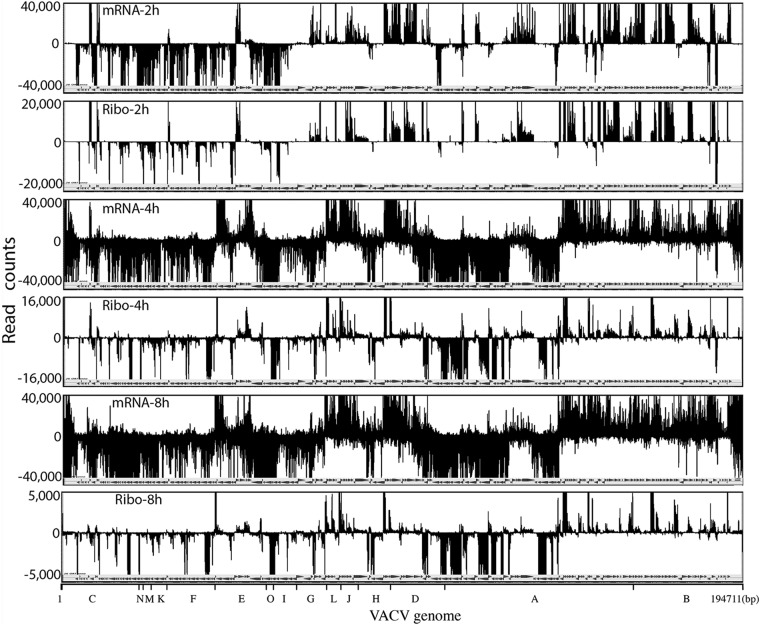
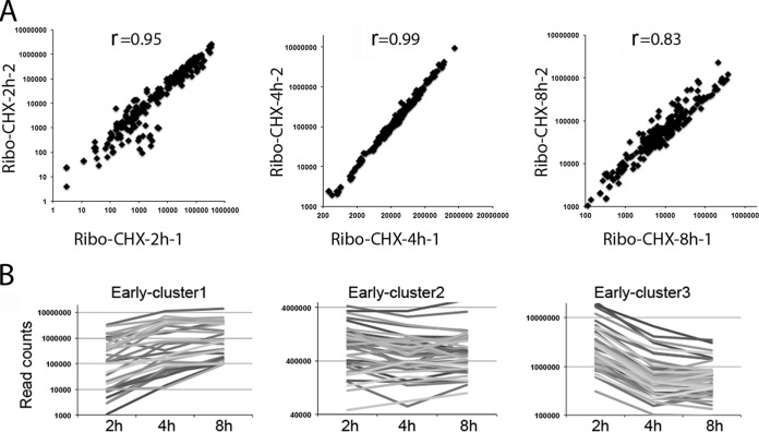
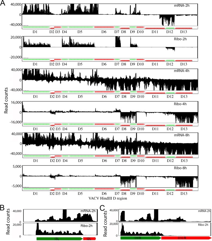
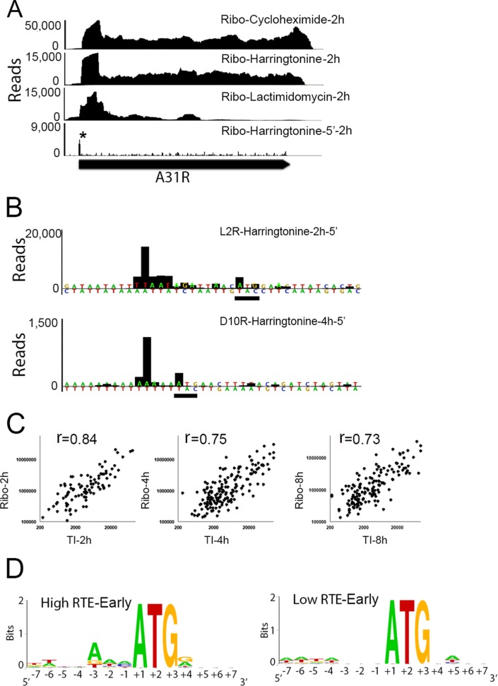
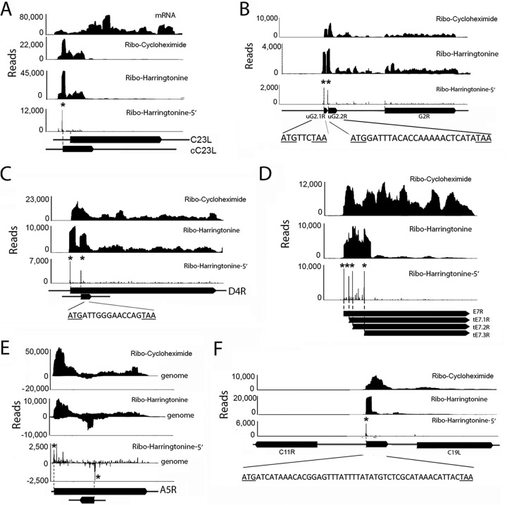
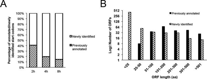
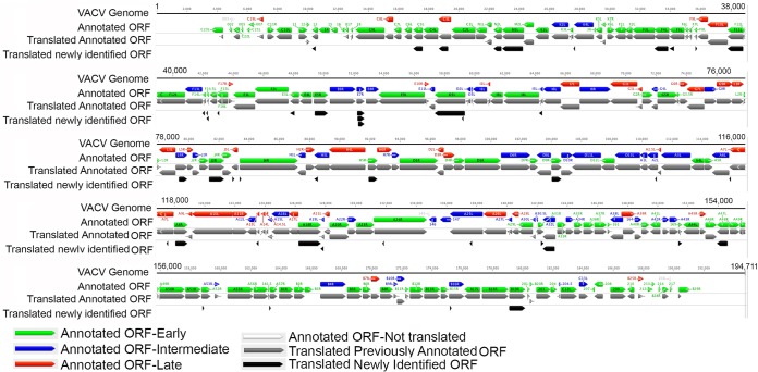
The more than 200 closely spaced annotated open reading frames, extensive transcriptional read-through, and numerous unpredicted RNA start sites have made the analysis of vaccinia virus gene expression challenging. Genome-wide ribosome profiling provided an unprecedented assessment of poxvirus gene expression. By 4 h after infection, approximately 80% of the ribosome-associated mRNA was viral. Ribosome-associated mRNAs were detected for most annotated early genes at 2 h and for most intermediate and late genes at 4 and 8 h. Cluster analysis identified a subset of early mRNAs that continued to be translated at the later times. At 2 h, there was excellent correlation between the abundance of individual mRNAs and the numbers of associated ribosomes, indicating that expression was primarily transcriptionally regulated. However, extensive transcriptional read-through invalidated similar correlations at later times. The mRNAs with the highest density of ribosomes had host response, DNA replication, and transcription roles at early times and were virion components at late times. Translation inhibitors were used to map initiation sites at single-nucleotide resolution at the start of most annotated open reading frames although in some cases a downstream methionine was used instead. Additional putative translational initiation sites with AUG or alternative codons occurred mostly within open reading frames, and fewer occurred in untranslated leader sequences, antisense strands, and intergenic regions. However, most open reading frames associated with these additional translation initiation sites were short, raising questions regarding their biological roles. The data were used to construct a high-resolution genome-wide map of the vaccinia virus translatome.

Importance: This report contains the first genome-wide, high-resolution analysis of poxvirus gene expression at both transcriptional and translational levels. The study was made possible by recent methodological advances allowing examination of the translated regions of mRNAs including start sites at single-nucleotide resolution. Vaccinia virus ribosome-associated mRNA sequences were detected for most annotated early genes at 2 h and for most intermediate and late genes at 4 and 8 h after infection. The ribosome profiling approach was particularly valuable for poxviruses because of the close spacing of approximately 200 open reading frames and extensive transcriptional read-through resulting in overlapping mRNAs. The expression of intermediate and late genes, in particular, was visualized with unprecedented clarity and quantitation. We also identified novel putative translation initiation sites that were mostly associated with short protein coding sequences. The results provide a framework for further studies of poxvirus gene expression.


Figures

FIG 1
FIG 1
Schematic diagram of the experimental approach. HeLa cells were infected with VACV WR at a multiplicity of 20 PFU/cell. Cycloheximide, harringtonine, or lactimidomycin was added at 2, 4, and 8 h postinfection, and mRNAs protected by ribosomes from digestion with RNase I were isolated and deep sequenced (ribosome profiling). Total mRNA in the presence of cycloheximide was simultaneously isolated, fragmented by digestion with RNase III, and deep sequenced (mRNA profiling).
FIG 2
FIG 2
Comparison of VACV genome-wide transcriptome and ribosome footprint maps at 2, 4, and 8 h of infection. The number of read counts per nucleotide is displayed over the genome-wide map of VACV ORFs. The reads above the line map to the upper DNA strand, and reads below the line map to the lower DNA strand. High read counts are off scale for display purpose. The VACV HindIII restriction endonuclease map is shown at the bottom. Abbreviations: mRNA, mRNA profiling; Ribo, ribosome profiling. The hours after infection are indicated.
FIG 3
FIG 3
Reproducibility and cluster analysis. (A) Ribosome profiling with cycloheximide (Ribo-CHX). Read counts were mapped to the annotated ORFs of VACV from two independent experiments at 2, 4, and 8 h postinfection. Each dot represents the read count for an individual mRNA. The correlation coefficients (r) are noted on each plot. (B) Cluster analysis of early gene expression. Cluster analysis was performed on the normalized read counts of ribosome footprints for each early gene at 2, 4, and 8 h after infection. Line plots of the read counts corresponding to three clusters are shown.
FIG 4
FIG 4
Comparison of mRNA and ribosome footprint reads in expanded regions of the VACV genome. (A) Transcription and ribosome footprint maps of the HindIII D region of the VACV genome at 2, 4, and 8 h of infection. The annotated ORFs D1 to D13 in this region are displayed, with green and red indicating early and postreplicative expression, respectively. (B and C) Transcripts and ribosome footprints corresponding to the I3L and D9R ORFs at 2 h of infection. Note that L and R following the ORF number indicate leftwards and rightwards transcription, respectively.
FIG 5
FIG 5
Identification of VACV translation initiation sites. (A) Ribosome footprints corresponding to A31R mRNA after treatment with cycloheximide, harringtonine, or lactimidomycin at 2 h of infection. The Ribo-harringtonine 5′ end shows only the first nucleotide of the ribosome footprint with the translation initiation site labeled by an asterisk. (B) Distance from the 5′ end of the ribosome footprint to the start codon for early L2R mRNA and late D10R mRNA. Multiple A residues were manually added to the sequence upstream of the D10R ORF to mimic the nontemplated A residues formed by RNA polymerase slippage. The ATG start codons are underlined. (C) Correlation between the number of reads associated with the translation initiation (TI) peak and the ribosome footprint (Ribo) for each individual mRNA indicated by a dot at 2, 4, and 8 h postinfection. (D) Logos of translation initiation sites and their surrounding nucleotides of the 20 early genes with the highest and the 20 early genes with the lowest relative translation efficiencies (RTE).
FIG 6
FIG 6
Representative novel translation units. Panels show mRNA reads and ribosome (Ribo)-protected reads aligned along portions of the VACV genome represented as filled arrows. Asterisks indicate the 5′ ends of the harringtonine peaks preceding the translation initiation sites. (A) RNA was obtained after adding cycloheximide or harringtonine at 2 h after infection, and the ribosome-protected reads were mapped along the C23L (VACWR001) ORF. The long filled arrow designated C23L is the annotated ORF. The short filled arrow designated cC23L represents the corrected ORF following the translation initiation site determined in this study (VACWR001). (B) Identification of translation initiation sites for two uORFs (uG2.1R and uG2.2R) upstream of the G2R (VACWR080) ORF. The sequences of the short ORFs are shown. (C) Identification of a translation initiation site for a downstream frameshifting ORF (dORF) in D4R (VACWR109). (D) Identification of translation initiation site for multiple truncated ORFs (tORFs) in E7R (VACWR062). (E) Identification of a translation initiation site for a novel ORF antisense to A5R (VACWR124). (F) Identification of a translation initiation site for an unannotated ORF between C19L (VACWR008) and C11R (VACWR009). The nucleotide sequence of the ORF is shown.
FIG 7
FIG 7
Comparison of the previously annotated and newly identified translation initiation sites. (A) Percentages of annotated and newly identified putative translation initiation sites at 2, 4, and 8 h of infection. (B) Size distribution of ORF lengths in amino acids (aa) associated with previously annotated and newly identified putative translation initiation sites.
FIG 8
FIG 8
VACV genome-wide expression map. The previously annotated ORFs of the WR strain of VACV are color coded as indicated and shown with arrows pointing in the direction of transcription. The annotated ORFs shown to be translated by ribosome profiling are indicated in gray. ORFs that start with AUG and are 25 amino acids or longer that were associated with new putative translation initiation sites are indicated in black. In many cases the additional new translation site is close to the previously annotated one so that the gray and black arrows appear similar in length.

References

    1. Moss B. 2013. Poxviridae, p 2129–2159. In Knipe DM, Howley PM, Cohen JI, Griffin DE, Lamb RA, Martin MA, Rancaniello VR, Roizman B (ed), Fields virology, 6th ed, vol 2 Lippincott Williams & Wilkins, Philadelphia, PA.
    1. Damon I. 2013. Poxviruses, p 2160–2184. In Knipe DM, Howley PM, Cohen JI, Griffin DE, Lamb RA, Martin MA, Rancaniello VR, Roizman B (ed), Fields virology, 6th ed, vol 2 Lippincott Williams & Wilkins, Philadelphia, PA.
    1. Wei CM, Moss B. 1975. Methylated nucleotides block 5′-terminus of vaccinia virus mRNA. Proc Natl Acad Sci U S A 72:318–322. doi:10.1073/pnas.72.1.318. - DOI - PMC - PubMed
    1. Martin SA, Paoletti E, Moss B. 1975. Purification of mRNA guanylyltransferase and mRNA (guanine 7-)methyltransferase from vaccinia virus. J Biol Chem 250:9322–9329. - PubMed
    1. Kates J, Beeson J. 1970. Ribonucleic acid synthesis in vaccinia virus. II. Synthesis of polyriboadenylic acid. J Mol Biol 50:19–23. - PubMed

Publication types

LinkOut - more resources