Review
. 2015 Apr 23;520(7548):466-73.
doi: 10.1038/nature14435.

A new heart for a new head in vertebrate cardiopharyngeal evolution

Affiliations
Review

A new heart for a new head in vertebrate cardiopharyngeal evolution

Rui Diogo et al. Nature. .

Abstract

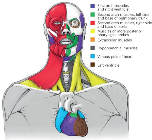
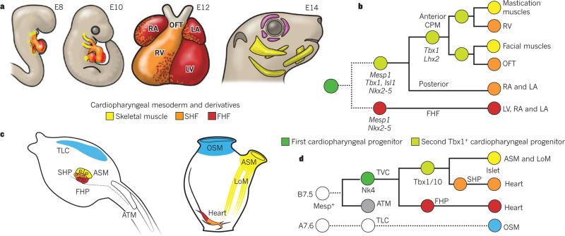
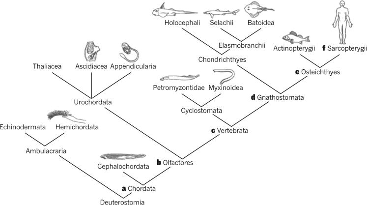
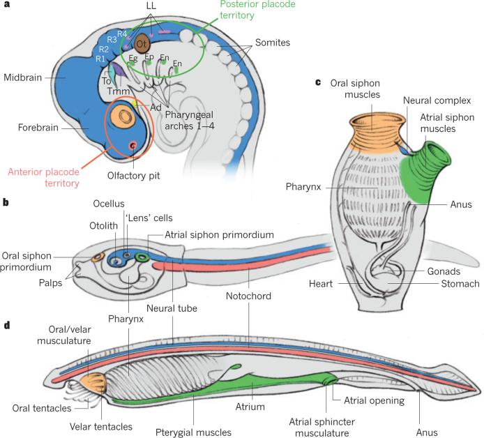
It has been more than 30 years since the publication of the new head hypothesis, which proposed that the vertebrate head is an evolutionary novelty resulting from the emergence of neural crest and cranial placodes. Neural crest generates the skull and associated connective tissues, whereas placodes produce sensory organs. However, neither crest nor placodes produce head muscles, which are a crucial component of the complex vertebrate head. We discuss emerging evidence for a surprising link between the evolution of head muscles and chambered hearts - both systems arise from a common pool of mesoderm progenitor cells within the cardiopharyngeal field of vertebrate embryos. We consider the origin of this field in non-vertebrate chordates and its evolution in vertebrates.


Figures

Figure 1
Figure 1. The striking heterogeneity of the human head and heart musculature
The head includes at least six different muscle groups, all arising from the cardiopharyngeal field and being branchiomeric, except the hypobranchial and perhaps the extraocular muscles. On the left side of the body (right part of figure) the facial expression muscles have been removed to show the masticatory muscles. The six groups are: first/mandibular arch muscles, including cells clonally related to the right ventricle; left second/hyoid arch muscles related to myocardium at the base of the pulmonary trunk; right second/hyoid arch muscles, related to myocardium at the base of the aorta; muscles of the most posterior pharyngeal arches, including muscles of the pharynx and larynx and the cucullaris-derived neck muscles trapezius and sternocleidomastoideus; extraocular muscles, which are often not considered to be branchiomeric, but according to classic embryological studies and recent retrospective clonal analyses in mice contain cells related to those of the branchiomeric mandibular muscles; and hypobranchial muscles, including tongue and infrahyoid muscles that derive from somites and migrate into the head and neck,,.
Figure 2
Figure 2. An evolutionarily conserved cardiopharyngeal ontogenetic motif
a, Mouse embryos at embryonic days (E)8 and 10, the four-chambered mouse heart at E12, and the mouse head at E14. First heart field (FHF)-derived regions of heart (left ventricle (LV) and atria) are in red; second heart field (SHF)-derived regions of heart (right ventricle (RV), left atrium (LA), right atrium (RA) and outflow tract (OFT)) are in orange; branchiomeric skeletal muscles are in yellow; extraocular muscles are in purple. b, Lineage tree depicting the origins of cardiac compartments and branchiomeric muscles in mice. All cells derive from common pan-cardiopharyngeal progenitors (dark green) that produce the FHF, precursors of the left ventricle and atria, and the second Tbx1+ cardiopharyngeal progenitors (light green). Broken lines indicate that the early common FHF and SHF progenitor remains to be identified in mice. In anterior cardiopharyngeal mesoderm (CPM), progenitor cells activate Lhx2, self-renew and produce the SHF-derived RV and OFT, and first and second arch branchiomeric muscles (including muscles of mastication and facial expression). c, Cardiopharyngeal precursors in Ciona intestinalis hatching larva (left) and their derivatives in the metamorphosed juvenile (right). The first heart precursors (FHP) (red) and second heart precursors (SHP) (orange) contribute to the heart (red and orange mix), whereas atrial siphon muscle precursors (ASM, yellow) form atrial siphon and longitudinal muscles (LoM, yellow). Oral siphon muscles (OSM, blue) derive from a heterogenous larval population of trunk lateral cells (TLC, blue). ATM, anterior tail muscles. CPM is bilaterally symmetrical around the midline (dotted line). d, Lineage tree depicting clonal relationships and gene activities deployed in C. intestinalis cardiopharyngeal precursors. All cells derive from Mesp+ B7.5 blastomeres, which produce ATM (grey, see also left panel of c) and trunk ventral cells (TVC, dark green). The latter pan-cardiopharyngeal progenitors express Nk4 and divide asymmetrically to produce the FHP (red) and second TVCs, the Tbx1/10+ second cardiopharyngeal progenitors (second TVC, light green disk). The latter divide again asymmetrically to produce SHP (orange) and the precursors of ASM and LoM, which upregulate Islet. The OSM arise from A7.6-derived trunk lateral cells (TLC, light blue).
Figure 3
Figure 3. Some of the synapomorphies of the Chordata and its subgroups, according to our own data and review of the literature
a, Somites and branchiomeric muscles. b, Placodes, neural-crest-like cells and cardiopharyngeal field (CPF) (although within invertebrates, conclusive evidence for these features was only reported in urochordates, some of these features may have been already present in the last common ancestor of extant chordates) giving rise to first- and second-heart-field-derived parts of the heart and to branchiomeric muscles (possibly not all of them, that is, inclusion of oral/velar muscles into CPF might have occurred during vertebrate evolution). c, Skull, cardiac chambers, and differentiation of epibranchial and hypobranchial somitic muscles. d, Jaws and differentiation between hypaxial and epaxial somitic musculature; paired appendages and fin muscles; origin of the branchiomeric muscle cucullaris. e, Loss of epibranchial muscles; cucullaris divided into levatores arcuum branchialium (going to pharyngeal arches) and protractor pectoralis (going to pectoral girdle), an exaptation that later allowed the emergence of the tetrapod neck. f, Within sarcopterygians, the protractor pectoralis gave rise to the amniote neck muscles trapezius and sternocleidomastoideus.
Figure 4
Figure 4. Homology hypotheses of placodes and branchiomeric muscles within chordates
a, Location of ectodermal placodes in the vertebrate head according to Graham and Shimeld's hypothesis (anterior to the left): olfactory placode or pit (red) at the tip of the forebrain; lens placodes (orange) form posteriorly as part of eye; adenohypophyseal placode (Ad, yellow) lies ventrally to forebrain; trigeminal placodes form alongside the anterior hindbrain at the levels of rhombomeres 1 and 2 (R1 and R2), the anterior one being the ophthalmic placode (To, light blue) and the posterior one the maxillomandibular placode (Tmm, purple); otic placode (Ot, brown) forms opposite the central domain of hindbrain; lateral line placodes (LL, pink) form anteriorly and posteriorly to otic placode; epibranchial placodes (green) — geniculate (Eg), petrosal (Ep) and nodose (En) — form as part of pharyngeal series. Forebrain, midbrain and R1–4, and neural tube are shown in dark blue. b, Urochordate tadpole-like larva (anterior to the left). The notochord is in red and two siphon primordia are in green and orange, with putative relationships to the anterior and posterior placode territories shown in a. c, Adult urochordate showing siphon primordia after metamorphosis. d, Adult cephalochordate showing the urochordate–cephalochordate muscle homology hypotheses proposed in the present Review. Figures based on images from refs , , .

References

    1. Gans C, Northcutt RG. Neural crest and the origin of vertebrates: a new head. Science. 1983;220:268–273. [This highly influential paper argued that the evolution of head structures derived from neural crest and cranial placodes had a crucial role in the transition to early vertebrates.] - PubMed
    1. Patthey C, Schlosser G, Shimeld SM. The evolutionary history of vertebrate cranial placodes — I: cell type evolution. Dev. Biol. 2014;389:82–97. - PubMed
    1. Graham A, Shimeld SM. The origin and evolution of the ectodermal placodes. J. Anat. 2013;222:32–40. - PMC - PubMed
    1. Northcutt RG. The new head hypothesis revisited. J. Exp. Zool. B Mol. Dev. Evol. 2005;304B:274–297. - PubMed
    1. Kuratani S. Evolution. A muscular perspective on vertebrate evolution. Science. 2013;341:139–140. - PubMed

Publication types