. 2015 Jun;123(2):205-16.
doi: 10.1007/s11060-015-1784-3. Epub 2015 Apr 23.

miRNA contents of cerebrospinal fluid extracellular vesicles in glioblastoma patients

Affiliations

miRNA contents of cerebrospinal fluid extracellular vesicles in glioblastoma patients

Johnny C Akers et al. J Neurooncol. 2015 Jun.

Abstract

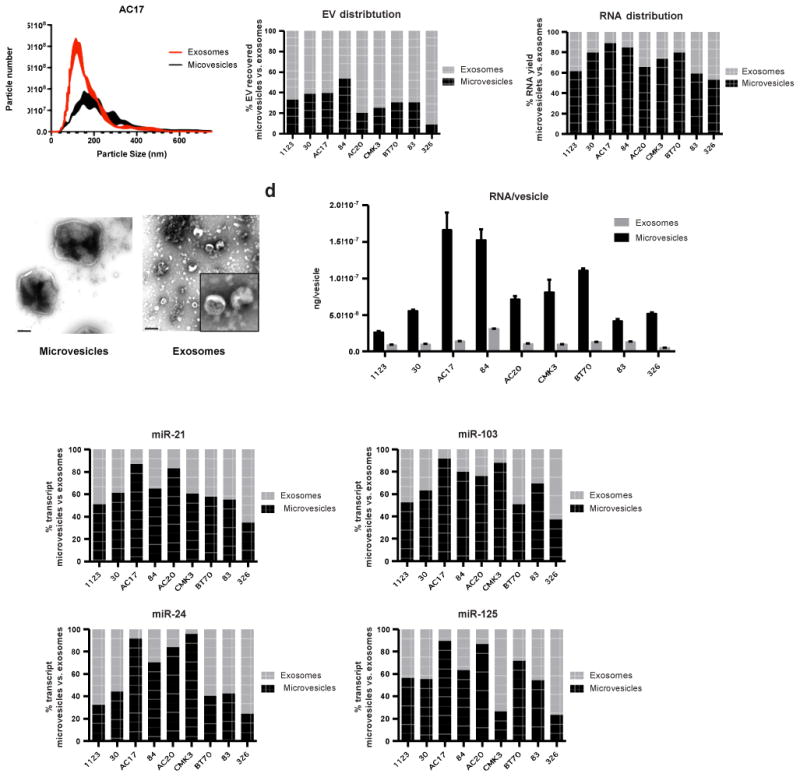
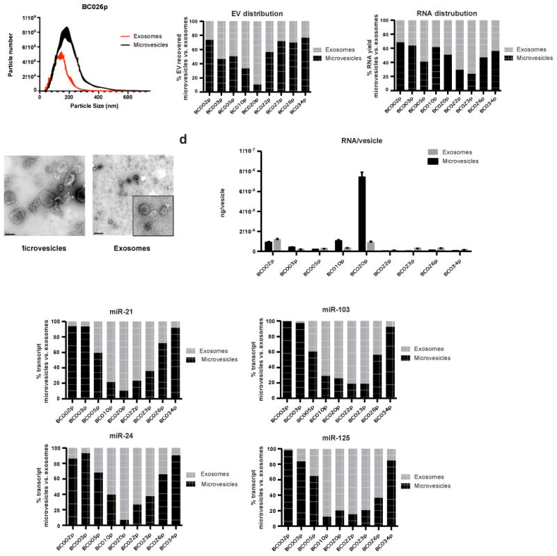
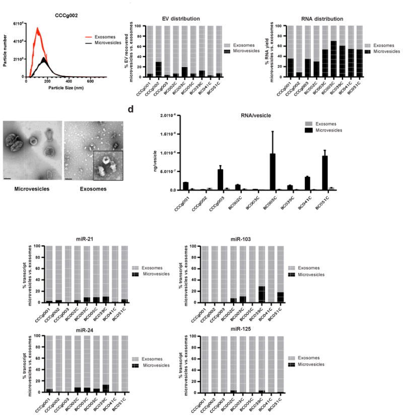
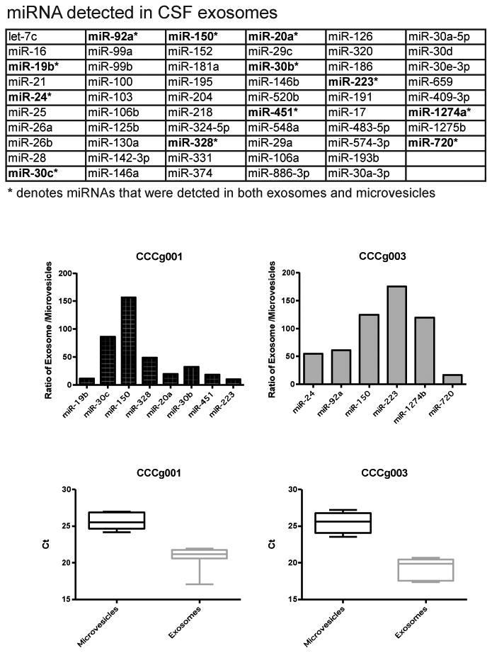
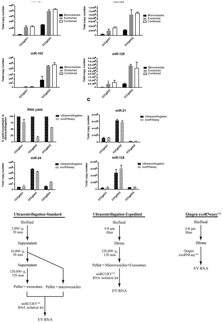
Analysis of extracellular vesicles (EVs) derived from plasma or cerebrospinal fluid (CSF) has emerged as a promising biomarker platform for therapeutic monitoring in glioblastoma patients. However, the contents of the various subpopulations of EVs in these clinical specimens remain poorly defined. Here we characterize the relative abundance of miRNA species in EVs derived from the serum and cerebrospinal fluid of glioblastoma patients. EVs were isolated from glioblastoma cell lines as well as the plasma and CSF of glioblastoma patients. The microvesicle subpopulation was isolated by pelleting at 10,000×g for 30 min after cellular debris was cleared by a 2000×g (20 min) spin. The exosome subpopulation was isolated by pelleting the microvesicle supernatant at 120,000×g (120 min). qRT-PCR was performed to examine the distribution of miR-21, miR-103, miR-24, and miR-125. Global miRNA profiling was performed in select glioblastoma CSF samples. In plasma and cell line derived EVs, the relative abundance of miRNAs in exosome and microvesicles were highly variable. In some specimens, the majority of the miRNA species were found in exosomes while in other, they were found in microvesicles. In contrast, CSF exosomes were enriched for miRNAs relative to CSF microvesicles. In CSF, there is an average of one molecule of miRNA per 150-25,000 EVs. Most EVs derived from clinical biofluids are devoid of miRNA content. The relative distribution of miRNA species in plasma exosomes or microvesicles is unpredictable. In contrast, CSF exosomes are the major EV compartment that harbor miRNAs.


Conflict of interest statement

Conflict of interest: None.

Figures

Fig. 1
Fig. 1. Distribution of select miRNAs in different fractions of glioblastoma cell line derived EVs
(a) Nanoparticle tracking analysis (NTA) of extracellular vesicles isolated from glioblastoma cell lines by differential centrifugation demonstrated distinct size profiles between microvesicles and exosomes. (b) Sizes of the isolated vesicles were confirmed by TEM. Scale bar represents 200 nm. Inset shows a magnified view of exosomes. (c) Contribution of microvesicle and exosome fractions to total EV and total RNA recovered. The relative abundance of microvesicles versus exosomes secreted by glioblastoma cells was variable across the nine lines. (d) Cell line-derived microvesicles contain more RNA per vesicle than the corresponding exosomes. Average RNA yield per vesicles was calculated by normalizing total RNA yield to vesicles number as determined by NTA. (e) The expression levels of miR-21, miR-103, miR-24, and miR-125 in microvesicles and exosomes were quantitatively assessed. qRT-PCR was performed in triplicate. All transcripts were detectable in both EV types, the distribution of each transcript among subpopulation of EVs were both transcript and cell line-dependent.
Fig. 2
Fig. 2. Distribution of select miRNAs in different fractions of glioblastoma patient plasma derived EVs
(a) Nanoparticle tracking analysis (NTA) of extracellular vesicles isolated from glioblastoma patient plasma by differential centrifugation. (b) Sizes of the isolated vesicles were confirmed by TEM. Scale bar represents 200 nm. Inset shows a magnified view of exosomes. (c) Contribution of microvesicle and exosome fractions to total EV and total RNA recovered. The relative abundance of microvesicles and exosomes in plasma also appeared to be highly variable across different clinical specimens. (d) Average RNA yield per vesicles isolated from plasma was calculated by normalizing total RNA yield to vesicles number as determined by NTA. In four out of nine patients' plasma, exosomes were found to contain higher RNA contents than microvesicles. (e) The expression levels miR-21, miR-103, miR-24, and miR-125 in microvesicles and exosomes were quantitatively assessed. qRT-PCR was performed in triplicate. All transcripts were detectable in both EV types, the distribution of each transcript among subpopulation of EVs were both transcript and specimen-dependent.
Fig. 3
Fig. 3. Distribution of select miRNAs in different fractions of glioblastoma patient cerebrospinal fluid (CSF) derived EVs
(a) Nanoparticle tracking analysis (NTA) of extracellular vesicles isolated from glioblastoma patient CSF by differential centrifugation. (b) Sizes of the isolated vesicles were confirmed by TEM. Scale bar represents 200 nm. Inset shows a magnified view of exosomes. (c) Contribution of microvesicle and exosome fractions to total EV and total RNA recovered in CSF. While the microvesicle fractions on average only accounted for 10.5% of the total number of CSF-derived EVs, due to the higher RNA content of microvesicles, ∼44.7% of total vesicular RNA recovered was extracted from these larger vesicles. (d) CSF derived microvesicles contain more RNA per vesicles than CSF derived exosomes. Average RNA yield per vesicles isolated from CSF was calculated by normalizing total RNA yield to vesicles number as determined by NTA. (e) The expression levels of miR-21, miR-103, miR-24, and miR-125 in microvesicles and exosomes were quantitatively assessed. qRT-PCR was performed in triplicate. In CSF, between 70.9 to 100% of the four miRNA transcripts assayed were found in the exosome fraction.
Fig. 4
Fig. 4. miRNA profiling of glioblastoma patient cerebrospinal fluid (CSF) derived EVs
RNAs isolated from CSF microvesices and exosomes were subjected to global miRNA profiling using the TaqMan OpenArray human microRNA panel. (a) List of miRNA detected in CSF exosomes. Asterisk denotes miRNA that were detectable in both CSF microvesicles and exosomes. (b) Of the 6-8 miRNAs that were detected in both exosomes and microvesicles, the relative abundance of these miRNAs were 3-150 fold higher in the exosomes relative to the microvesicles. (c) On average, these miRNAs were detected 5-7 CT values lower in exosomes relative to microvesicles. These results suggest that miRNAs, in general, are enriched in the CSF exosomal fractions.
Fig. 5
Fig. 5. Streamlined method for CSF exosome isolation
(a) CSF EVs were isolated using differential centrifugation as described into microvesicle and exosome fractions or pelleted in a single 120,000×g spin (labeled as combined. For each of the four transcripts tested, similar levels of RNA were detected in both exosome pellet and the combined microvesicle+exosome pellet. (b) Evaluation of exoRNeasy Maxi Kit by Qiagen for the isolation of RNA from CSF EVs. Comparing total RNA yield, ultracentrifugation followed by RNA extraction using the Exiqon miRCURY RNA isolation kit recovered 1.3 to 5.5-fold more nucleic acid than the exoRNeasy kit. (c) The total copy number of miR-21, miR-24, and miR-125 recovered by ultracentrifugation or exoRNeasy was quantitatively assessed. qRT-PCR was performed in triplicate. Similar amount of miRNAs were recovered by ultracentrifugation and exoRNeasy. (d) Schematic representation of protocol used for the isolation of CSF EV RNA.

References

    1. Ostrom QT, Gittleman H, Farah P, Ondracek A, Chen Y, et al. CBTRUS statistical report: Primary brain and central nervous system tumors diagnosed in the United States in 2006-2010. Neuro Oncol. 2013;15(Suppl 2):ii1–56. - PMC - PubMed
    1. Bartek J, Jr, Ng K, Bartek J, Fischer W, Carter B, et al. Key concepts in glioblastoma therapy. J Neurol Neurosurg Psychiatry. 2012;83:753–760. - PubMed
    1. Stupp R, Hegi ME, Mason WP, van den Bent MJ, Taphoorn MJ, et al. Effects of radiotherapy with concomitant and adjuvant temozolomide versus radiotherapy alone on survival in glioblastoma in a randomised phase III study: 5-year analysis of the EORTC-NCIC trial. Lancet Oncol. 2009;10:459–466. - PubMed
    1. Clarke JL, Chang SM. Neuroimaging: diagnosis and response assessment in glioblastoma. Cancer J. 2012;18:26–31. - PubMed
    1. Sorensen AG, Batchelor TT, Wen PY, Zhang WT, Jain RK. Response criteria for glioma. Nat Clin Pract Oncol. 2008;5:634–644. - PMC - PubMed

Publication types

MeSH terms