

A review of neuroimaging findings in repetitive brain trauma
- PMID: 25904047
- PMCID: PMC5699448
- DOI: 10.1111/bpa.12249
A review of neuroimaging findings in repetitive brain trauma
Abstract
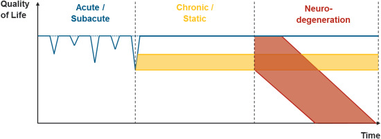
Chronic traumatic encephalopathy (CTE) is a neurodegenerative disease confirmed at postmortem. Those at highest risk are professional athletes who participate in contact sports and military personnel who are exposed to repetitive blast events. All neuropathologically confirmed CTE cases, to date, have had a history of repetitive head impacts. This suggests that repetitive head impacts may be necessary for the initiation of the pathogenetic cascade that, in some cases, leads to CTE. Importantly, while all CTE appears to result from repetitive brain trauma, not all repetitive brain trauma results in CTE. Magnetic resonance imaging has great potential for understanding better the underlying mechanisms of repetitive brain trauma. In this review, we provide an overview of advanced imaging techniques currently used to investigate brain anomalies. We also provide an overview of neuroimaging findings in those exposed to repetitive head impacts in the acute/subacute and chronic phase of injury and in more neurodegenerative phases of injury, as well as in military personnel exposed to repetitive head impacts. Finally, we discuss future directions for research that will likely lead to a better understanding of the underlying mechanisms separating those who recover from repetitive brain trauma vs. those who go on to develop CTE.
Keywords: neuroimaging; repetitive head injury.
© 2015 International Society of Neuropathology.
Figures
References
-
- Abbas K, Shenk TE, Poole VN, Breedlove EL, Leverenz LJ, Nauman EA et al (2014) Alteration of default mode network in high school football athletes due to repetitive subconcussive mild traumatic brain injury: a resting‐state functional magnetic resonance imaging study. Brain Connect. - PubMed
-
- Agdeppa ED, Kepe V, Liu J, Flores‐Torres S, Satyamurthy N, Petric A et al (2001) Binding characteristics of radiofluorinated 6‐dialkylamino‐2‐naphthylethylidene derivatives as positron emission tomography imaging probes for beta‐amyloid plaques in Alzheimer's disease. J Neurosci 21:RC189. - PMC - PubMed
-
- Albaugh MD, Orr C, Nickerson JP, Zweber C, Slauterbeck JR, Hipko S et al (2015) Postconcussive symptoms are associated with cerebral cortical thickness in healthy collegiate and preparatory school ice hockey players. J Pediatr 166:394–400. - PubMed
-
- Amen DG, Wu JC, Taylor D, Willeumier K (2011) Reversing brain damage in former NFL players: implications for traumatic brain injury and substance abuse rehabilitation. J Psychoactive Drugs 43:1–5. - PubMed
Publication types
MeSH terms
Grants and funding
LinkOut - more resources
Full Text Sources
Other Literature Sources
Research Materials
