. 2015 Jun;26(6):974-85.
doi: 10.1007/s13361-015-1147-5. Epub 2015 Apr 23.

MALDI FTICR IMS of Intact Proteins: Using Mass Accuracy to Link Protein Images with Proteomics Data

Affiliations

MALDI FTICR IMS of Intact Proteins: Using Mass Accuracy to Link Protein Images with Proteomics Data

Jeffrey M Spraggins et al. J Am Soc Mass Spectrom. 2015 Jun.

Abstract

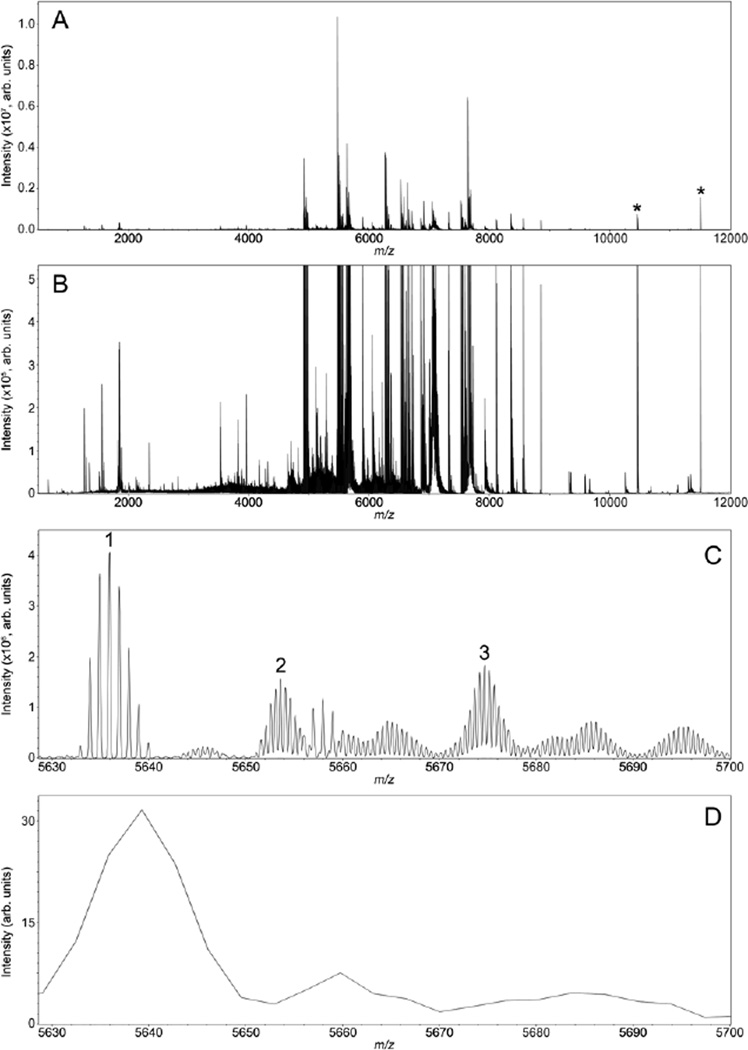
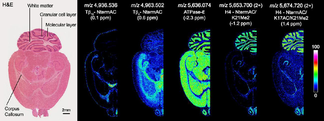
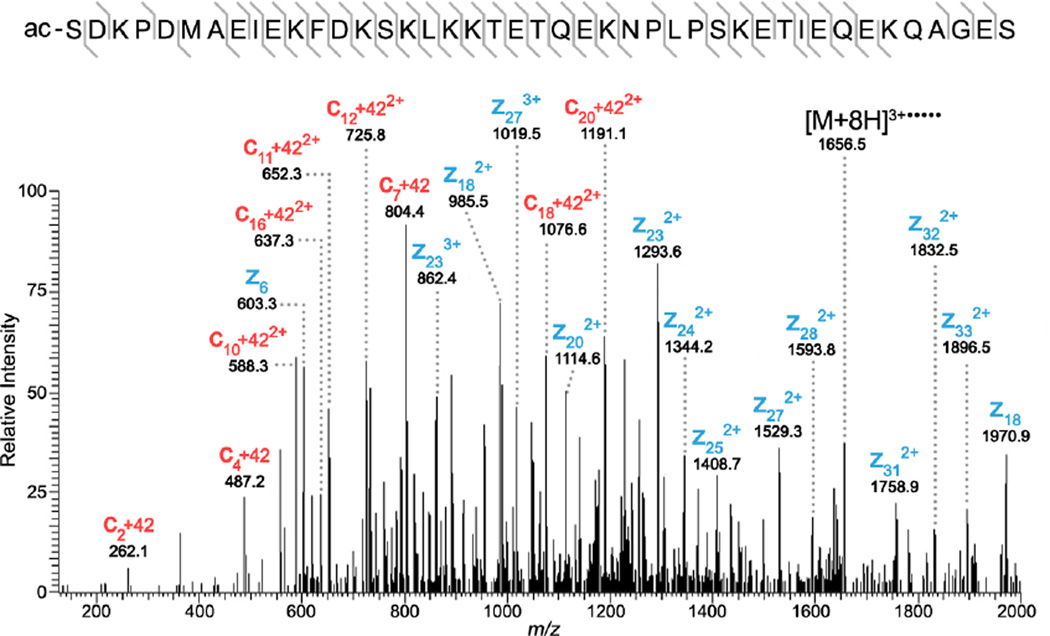
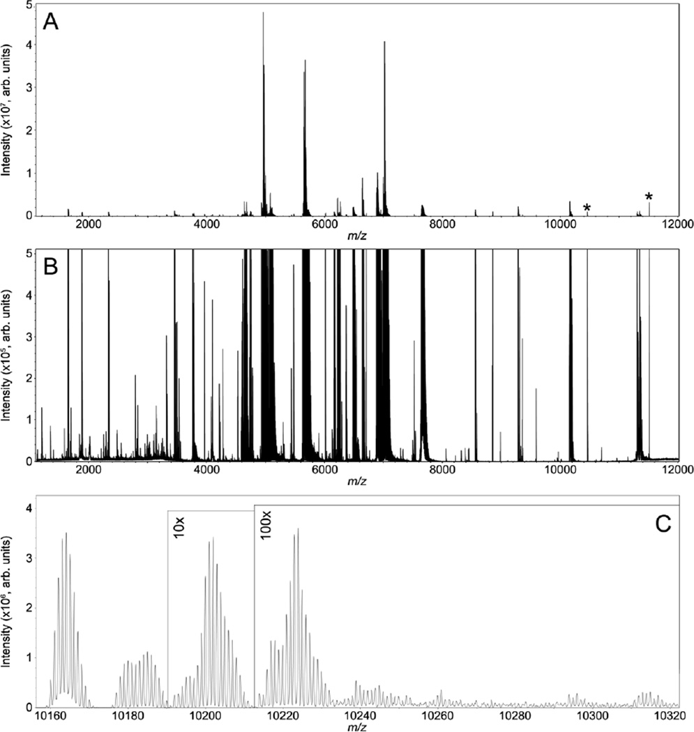
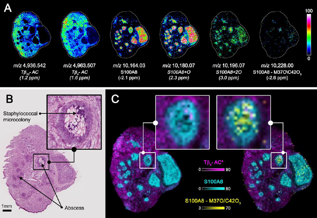
MALDI imaging mass spectrometry is a highly sensitive and selective tool used to visualize biomolecules in tissue. However, identification of detected proteins remains a difficult task. Indirect identification strategies have been limited by insufficient mass accuracy to confidently link ion images to proteomics data. Here, we demonstrate the capabilities of MALDI FTICR MS for imaging intact proteins. MALDI FTICR IMS provides an unprecedented combination of mass resolving power (~75,000 at m/z 5000) and accuracy (<5ppm) for proteins up to ~12kDa, enabling identification based on correlation with LC-MS/MS proteomics data. Analysis of rat brain tissue was performed as a proof-of-concept highlighting the capabilities of this approach by imaging and identifying a number of proteins including N-terminally acetylated thymosin β(4) (m/z 4,963.502, 0.6ppm) and ATP synthase subunit ε (m/z 5,636.074, -2.3ppm). MALDI FTICR IMS was also used to differentiate a series of oxidation products of S100A8 (m/z 10,164.03, -2.1ppm), a subunit of the heterodimer calprotectin, in kidney tissue from mice infected with Staphylococcus aureus. S100A8 - M37O/C42O(3) (m/z 10228.00, -2.6ppm) was found to co-localize with bacterial microcolonies at the center of infectious foci. The ability of MALDI FTICR IMS to distinguish S100A8 modifications is critical to understanding calprotectin's roll in nutritional immunity.


Figures

Figure 1
Figure 1
MALDI FTICR IMS of intact proteins from rat brain tissue. The average spectrum of the entire imaging data set is shown in panel A. Expanding the intensity scale (B) highlights the overall complexity and quality of the data with singly and doubly charged protein signals detected between m/z 1,000 – 12,000. Data were collected with a resolving power of ∼40,000 at m/z 5,000 providing isotopic resolution and allowing ions of different charge states and modifications to be distinguished (Panel C). The ion labeled 1 is singly charged and ions labeled 2 and 3 are examples of doubly charged ions. For comparison MALDI TOF IMS data set collected in linear mode from a serial tissue section is displayed in Panel D. Electronic noise peaks are labeled ( * ).
Figure 2
Figure 2
Selected ion images of intact proteins from rat brain tissue collected using MALDI FTICR MS. Ions were identified using mass accuracy to correlate imaging results with separate top-down proteomics experiments. For comparison, the tissue was H&E stained following IMS analysis. Doubly charged ions are indicated.
Figure 3
Figure 3
High mass resolution top-down ETD for the [M+8H]8+ charge state of thymosin β4. Selected c (red) and z (blue) ions are labeled clearly showing acetylation on the N-terminus. The amino acid sequence highlighting the fragmentation coverage is also provided.
Figure 4
Figure 4
MALDI FTICR IMS of intact proteins from kidney tissue from a mouse infected with S. aureus. The average spectrum of the entire imaging data set is shown in panel A. Expanding the intensity scale (B) highlights the overall quality of the data with singly and doubly charged protein signals detected between m/z 1,000 - 12,000. Data were collected with a resolving power of ∼75,000 at m/z 5,000 providing isotopic resolution and allowing multiple isoforms of S100A8 to be distinguished (Panel C). Electronic noise peaks are labeled (*).
Figure 5
Figure 5
Selected ion images of intact proteins from kidney tissue from a mouse infected with Staphylococcus aureus collected using MALDI FTICR MS (A). Ions were identified using mass accuracy to correlate imaging results with separate top-down proteomics experiments. For comparison, a serial tissue section was H&E stained (B). Ion image overlays show the advance oxidation product S100A8 – M7O/C42O3 is localized specifically to the center of infectious foci (C). Italicized ions were tentatively identified by mass accuracy only.
Figure 6
Figure 6
LC-MS/MS results for S100A8 – M37O/C42O3. Panel A shows high mass resolution top-down ETD data for the [M+12H]12+ parent ion with selected c (red) and z (blue) ions labeled. The amino acid sequence summarizing both top-down and bottom-up fragmentation coverage is provided (B). Grey fragment ion indicators designate top-down ions and pink indicators represent bottom-up results. Panel C shows high mass resolution bottom-up fragmentation data for the tryptic peptide highlighted in pink (B). The resulting fragments clearly show oxidation to both methionine 37 (+O) and cysteine 42 (+3O).

References

    1. Caprioli RM, Farmer TB, Gile J. Molecular imaging of biological samples: localization of peptides and proteins using MALDI-TOF MS. Anal Chem. 1997;69:4751–4760. - PubMed
    1. Nilsson A, Goodwin RJ, Shariatgorji M, Vallianatou T, Webborn PJ, Andren PE. Mass spectrometry imaging in drug development. Anal Chem. 2015;87:1437–1455. - PubMed
    1. Huang JT, Hannah-Qiuhua L, Szyszka R, Veselov V, Reed G, Wang X, et al. Molecular imaging of drug-eluting coronary stents: method development, optimization and selected applications. J Mass Spectrom. 2012;47:155–162. - PubMed
    1. Bhandari DR, Schott M, Rompp A, Vilcinskas A, Spengler B. Metabolite localization by atmospheric pressure high-resolution scanning microprobe matrix-assisted laser desorption/ionization mass spectrometry imaging in whole-body sections and individual organs of the rove beetle Paederus riparius. Anal Bioanal Chem. 2014 - PMC - PubMed
    1. Berry KA, Hankin JA, Barkley RM, Spraggins JM, Caprioli RM, Murphy RC. MALDI imaging of lipid biochemistry in tissues by mass spectrometry. Chem Rev. 2011;111:6491–6512. - PMC - PubMed

Publication types

MeSH terms

LinkOut - more resources