. 2015 Jun;21(6):1203-16.
doi: 10.1261/rna.049171.114. Epub 2015 Apr 22.

The C-terminal helix in the YjeQ zinc-finger domain catalyzes the release of RbfA during 30S ribosome subunit assembly

Affiliations

The C-terminal helix in the YjeQ zinc-finger domain catalyzes the release of RbfA during 30S ribosome subunit assembly

Ajitha Jeganathan et al. RNA. 2015 Jun.

Abstract

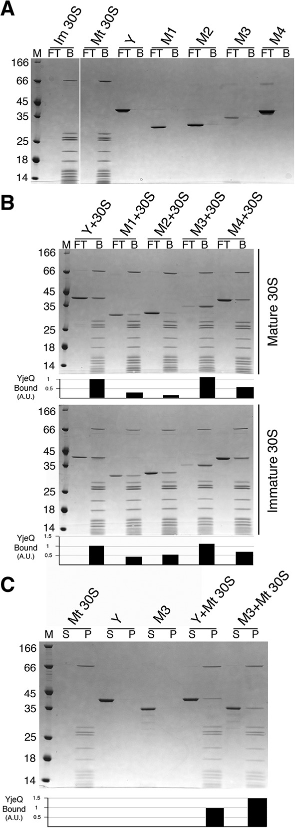
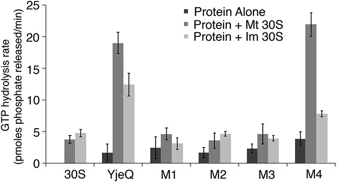
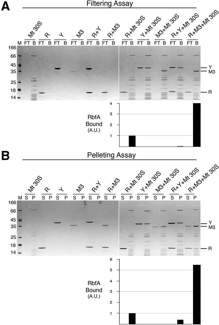
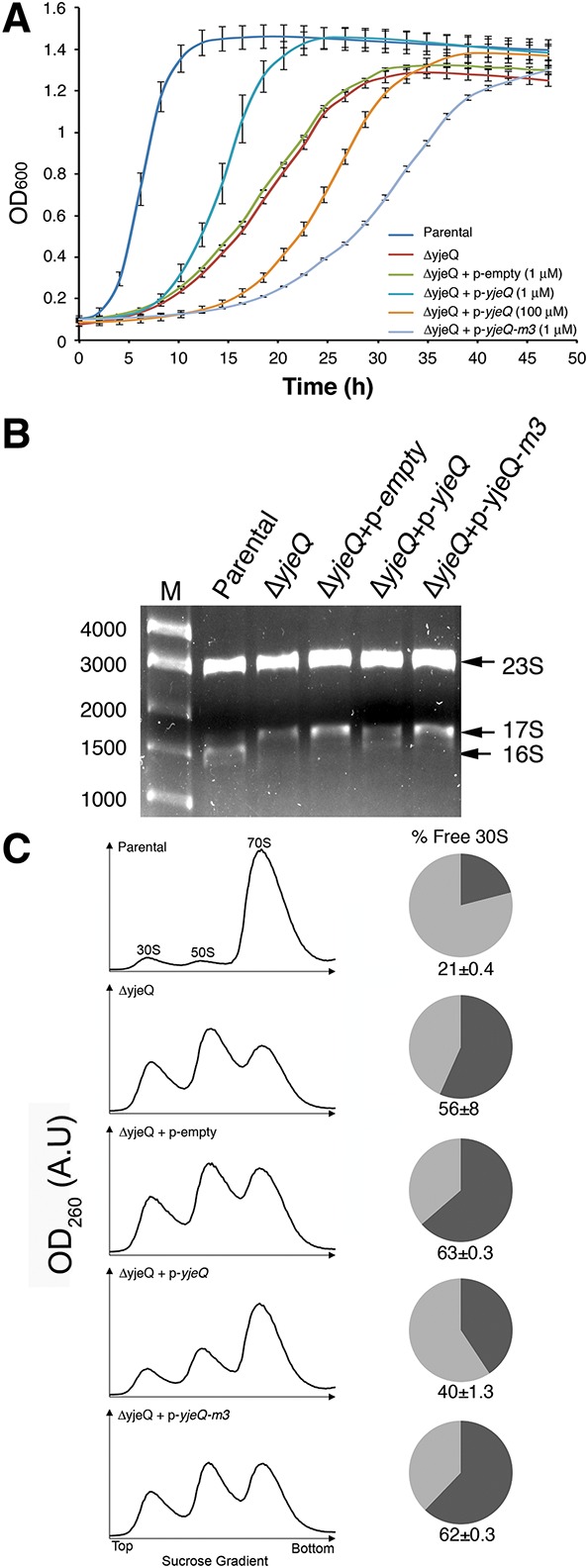
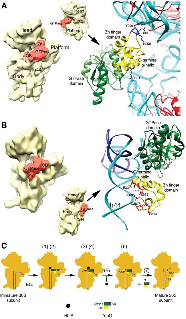
YjeQ (also called RsgA) and RbfA proteins in Escherichia coli bind to immature 30S ribosome subunits at late stages of assembly to assist folding of the decoding center. A key step for the subunit to enter the pool of actively translating ribosomes is the release of these factors. YjeQ promotes dissociation of RbfA during the final stages of maturation; however, the mechanism implementing this functional interplay has not been elucidated. YjeQ features an amino-terminal oligonucleotide/oligosaccharide binding domain, a central GTPase module and a carboxy-terminal zinc-finger domain. We found that the zinc-finger domain is comprised of two functional motifs: the region coordinating the zinc ion and a carboxy-terminal α-helix. The first motif is essential for the anchoring of YjeQ to the 30S subunit and the carboxy-terminal α-helix facilitates the removal of RbfA once the 30S subunit reaches the mature state. Furthermore, the ability of the mature 30S subunit to stimulate YjeQ GTPase activity also depends on the carboxy-terminal α-helix. Our data are consistent with a model in which YjeQ uses this carboxy-terminal α-helix as a sensor to gauge the conformation of helix 44, an essential motif of the decoding center. According to this model, the mature conformation of helix 44 is sensed by the carboxy-terminal α-helix, which in turn stimulates the YjeQ GTPase activity. Hydrolysis of GTP is believed to assist the release of YjeQ from the mature 30S subunit through a still uncharacterized mechanism. These results identify the structural determinants in YjeQ that implement the functional interplay with RbfA.

Keywords: 30S subunit; GTPase; RbfA protein; RsgA protein; YjeQ protein; ribosome assembly.


Figures

FIGURE 1.
FIGURE 1.
Binding of YjeQ carboxy-terminal variants to the mature and immature 30S subunits. (A,B) Ability of YjeQ (Y) and YjeQ variants (M1, M2, M3, and M4) to bind to the mature (Mt) and immature (Im) 30S subunits analyzed by filtration assays. Coomassie blue stained SDS-PAGE in A contains the controls for the experiment consistent in reactions containing either YjeQ (full-length or variants) or 30S subunits (mature or immature) by themselves. Reactions containing a mixture of YjeQ protein and ribosomal particles contained a fivefold molar excess of protein. Assembly mixtures were incubated for 15 min at 16°C in the presence of 1 mM GMP-PNP. Following incubation, the reactions were passed through a 100 kDa cut-off filter using centrifugation. The unbound protein was captured in the flow-through (FT) and the protein bound to the ribosomal particles (B) was retained by the filter and resuspended by an equal volume of buffer. The molecular weight marker (M) is in kDa. The flow-through and bound portions from these assays were loaded into 4%–12% bis–tris polyacrylamide gels and resolved using SDS-PAGE. (C) Pelleting assay of YjeQ M3 variant with the mature 30S subunit. A fivefold excess of YjeQ M3 was incubated with mature 30S subunits for 15 min at 16°C. Following the incubation, reactions were laid over a sucrose cushion and subjected to ultracentrifugation. Proteins that were unbound were collected in the supernatant (S), while proteins that bound to the 30S particle were found in the pellet (P). The molecular weight (M) is in kDa. The pellet and supernatant were resolved by 4%–12% bis–tris SDS-PAGE and stained with Coomassie blue. The bar diagrams under the gels in B and C indicate the binding of the YjeQ variants to the 30S subunits with respect to wild-type YjeQ (set as 1).
FIGURE 2.
FIGURE 2.
Stimulation of GTPase activity of YjeQ variants by mature and immature 30S subunits. The GTP hydrolysis rate of the YjeQ variants alone or in the presence of mature (Mt 30S) and immature 30S (Im 30S) subunits was assessed using the Malachite Green Phosphate assay as described in Materials and Methods. The GTPase hydrolysis rates plotted in the graph were determined by measuring the free phosphate produced after the reactions had been incubated for 60 min at 25°C. Standard deviations shown in the plot correspond to three replicas of the experiment.
FIGURE 3.
FIGURE 3.
YjeQ M3 is unable to remove RbfA bound to the mature 30S subunit. (A) Filtration assay to test the release of RbfA (R) from the mature 30S subunit (Mt 30S) in the presence of YjeQ wild-type (Y) and YjeQ M3 variant (M3). These reactions were performed by adding the assembly factors in fivefold molar excess with respect to the 30S subunits and in the presence of GMP-PNP. Incubations were done at 25°C. Flow-through (FT) and bound (B) fractions from the filtration assay were resolved in 4%–12% bis–tris SDS-PAGE and stained with Coomassie blue. (B) Pelleting assay of identical reactions used in A. The molecular weight marker (M) is in kDa. Supernatant (S) and pellet (P) fractions from this assay were resolved in 4%–12% bis–tris SDS-PAGE and stained with Coomassie blue. The bar diagrams under the gels indicate the binding of the RbfA to the mature 30S subunit in each reaction. The observed binding of RbfA to the mature 30S subunit was defined as 1.
FIGURE 4.
FIGURE 4.
The carboxy-terminal extension of the zinc-finger domain in YjeQ is necessary for its function in vivo. (A) The parental strain and the yjeQ null strain by itself or complemented with the plasmid expressing yjeQ, YjeQ M3 variant, or the empty vector were induced at t = 0 h with the indicated concentration of IPTG and grown at 25°C for 47 h. Growth was monitored by measuring absorbance at 600 nm and plotted against time. Standard deviations shown in the plot correspond to three replicas of the experiment. (B) Total rRNA of cell cultures were analyzed when cultures reached mid-log phase represented by OD600 = 0.2. Total rRNA was extracted and resolved by electrophoresis in 0.9% synergel–0.7% agarose gel. The marker (M) is in base pairs. (C) Ribosome absorbance profiles from the parental strain and yjeQ null strain by itself or complemented with the plasmid expressing YjeQ, YjeQ M3 variant, or the empty plasmid were fractionated by ultracentrifugation in 10%–30% sucrose gradients providing the profiles shown in this panel. Peaks for the 30S, 50S subunits, and 70S ribosomes are indicated. The proportion of free 30S to bound 30S (i.e., 30S subunits in 70S complexes) in each case was calculated by integrating the areas under the 30S and 70S peaks of the sucrose gradient profiles. The area of the 30S peak plus one-third the area of the 70S peak corresponds to the total 30S population. The area of the 30S peak was divided by the total 30S absorbance to obtain the percentage of free 30S subunits and produce the pie charts in this panel. The standard deviations shown correspond to three replicas of the experiment. Peak area for the 30S subunit in each case was measured with respect to the area under the 70S peak to calculate the percentage of free 30S subunits in both strains. The calculated percentages are shown in the pie charts. The standard deviations shown correspond to three replicas of the experiment.
FIGURE 5.
FIGURE 5.
Interaction of the zinc-finger domain of YjeQ with the 30S subunit and working model for the functional interplay between YjeQ and RbfA. Structures of the 30S+YjeQ complex as described in Guo et al. (2011) (A) and Jomaa et al. (2011b) (B). The panel in the left shows a view of the entire complex. Landmarks and domains of the YjeQ protein are labeled. Area colored in red represents the YjeQ protein bound to the 30S subunit. The panel in the right shows a zoomed in view of the complex in the area of interaction of the zinc-finger domain with the 30S subunit. The rRNA is colored in cyan except for some nucleotides (B) that are colored in dark blue. A corresponding density for these nucleotides does not exist in the cryo-EM map of the 30S+YjeQ complex (Jomaa et al. 2011b). The side chains of important residues for this interaction are labeled. Structures shown were obtained from the EMDB (EMD-1884 and EMD-1895) and PDB (2YKR and 4A2I). Images of the structures were prepared with UCSF Chimera software (Pettersen et al. 2004). (C) Proposed model for the functional interplay between YjeQ and RbfA. The model explains the mechanism through which YjeQ facilitates the release of RbfA once the maturation of the 30S subunit is completed. Numbers in brackets refer to the steps of the model described in the text.

References

    1. Anand B, Verma SK, Prakash B 2006. Structural stabilization of GTP-binding domains in circularly permuted GTPases: implications for RNA binding. Nucleic Acids Res 34: 2196–2205. - PMC - PubMed
    1. Baba T, Ara T, Hasegawa M, Takai Y, Okumura Y, Baba M, Datsenko KA, Tomita M, Wanner BL, Mori H 2006. Construction of Escherichia coli K-12 in-frame, single-gene knockout mutants: the Keio collection. Mol Syst Biol 2: 2006.0008. - PMC - PubMed
    1. Ban N, Nissen P, Hansen J, Moore PB, Steitz TA 2000. The complete atomic structure of the large ribosomal subunit at 2.4 Å resolution. Science 289: 905–920. - PubMed
    1. Boehringer D, O'Farrell HC, Rife JP, Ban N 2012. Structural insights into methyltransferase KsgA function in 30S ribosomal subunit biogenesis. J Biol Chem 287: 10453–10459. - PMC - PubMed
    1. Bylund GO, Wipemo LC, Lundberg LA, Wikstrom PM 1998. RimM and RbfA are essential for efficient processing of 16S rRNA in Escherichia coli. J Bacteriol 180: 73–82. - PMC - PubMed

Publication types

MeSH terms

LinkOut - more resources