. 2015 Jul;114(1):400-10.
doi: 10.1152/jn.00136.2015. Epub 2015 Apr 22.

Periodic modulation of repetitively elicited monosynaptic reflexes of the human lumbosacral spinal cord

Affiliations

Periodic modulation of repetitively elicited monosynaptic reflexes of the human lumbosacral spinal cord

Ursula S Hofstoetter et al. J Neurophysiol. 2015 Jul.

Abstract

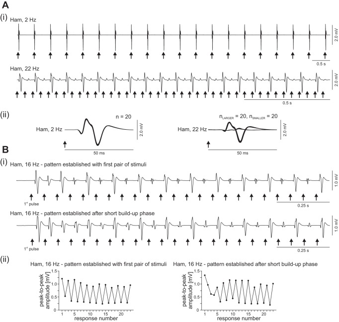
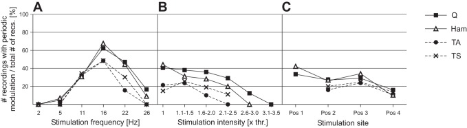
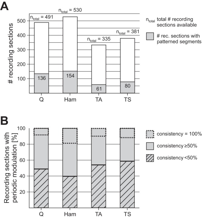
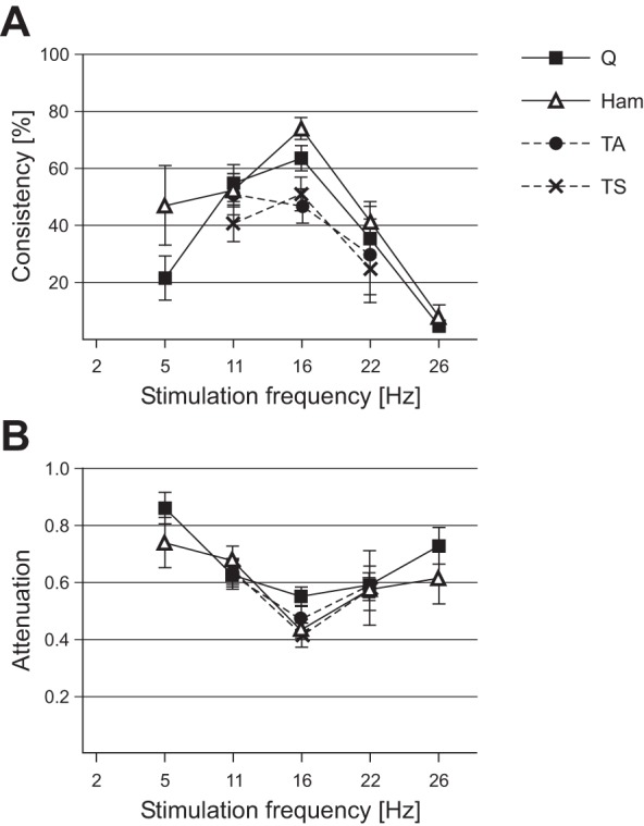
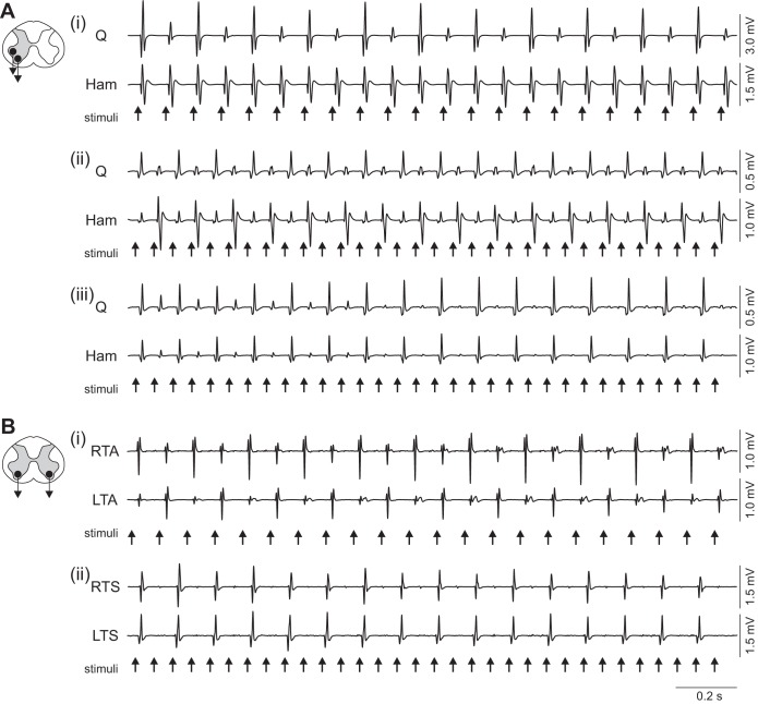
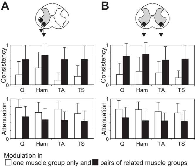
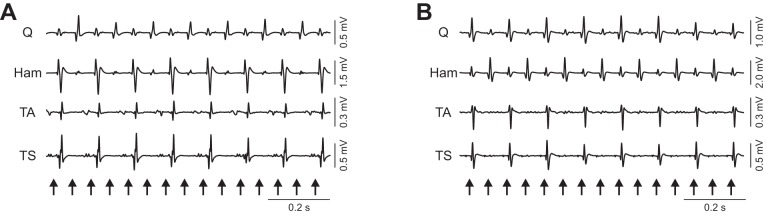
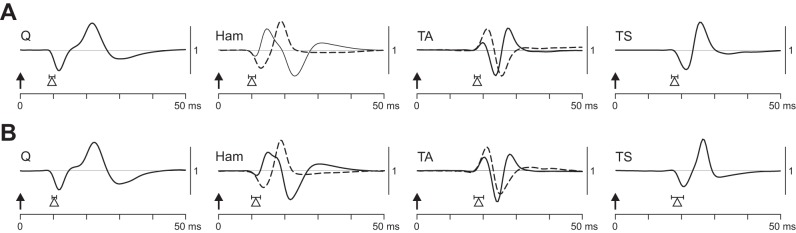
In individuals with motor-complete spinal cord injury, epidural stimulation of the lumbosacral spinal cord at 2 Hz evokes unmodulated reflexes in the lower limbs, while stimulation at 22-60 Hz can generate rhythmic burstlike activity. Here we elaborated on an output pattern emerging at transitional stimulation frequencies with consecutively elicited reflexes alternating between large and small. We analyzed responses concomitantly elicited in thigh and leg muscle groups bilaterally by epidural stimulation in eight motor-complete spinal cord-injured individuals. Periodic amplitude modulation of at least 20 successive responses occurred in 31.4% of all available data sets with stimulation frequency set at 5-26 Hz, with highest prevalence at 16 Hz. It could be evoked in a single muscle group only but was more strongly expressed and consistent when occurring in pairs of antagonists or in the same muscle group bilaterally. Latencies and waveforms of the modulated reflexes corresponded to those of the unmodulated, monosynaptic responses to 2-Hz stimulation. We suggest that the cyclical changes of reflex excitability resulted from the interaction of facilitatory and inhibitory mechanisms emerging after specific delays and with distinct durations, including postactivation depression, recurrent inhibition and facilitation, as well as reafferent feedback activation. The emergence of large responses within the patterns at a rate of 5.5/s or 8/s may further suggest the entrainment of spinal mechanisms as involved in clonus. The study demonstrates that the human lumbosacral spinal cord can organize a simple form of rhythmicity through the repetitive activation of spinal reflex circuits.

Keywords: human; repetitive nerve stimulation; rhythm generation; spinal cord stimulation; spinal reflexes.


Figures

Fig. 1.
Fig. 1.
Series of posterior root-muscle (PRM) reflexes without and with periodic amplitude modulation elicited by epidural stimulation of the lumbosacral spinal cord. Ai, top: responses of hamstrings (Ham) to 2-Hz stimulation with unmodulated amplitudes. Bottom: periodic reflex modulation elicited at 22 Hz in the same muscle group (note different time scaling). ii: Same PRM reflexes as shown in i but presented stimulus triggered in enlarged timescale. n, Numbers of superimposed responses. Bi: examples illustrating the characteristic short buildup phase of periodic modulation patterns, here elicited at 16 Hz, established either with the first pair of stimuli applied (top) or after a few stimulus pulses (bottom). ii: Peak-to-peak amplitudes (black circles) of the consecutively elicited PRM reflexes shown in i. Arrows mark times of stimulus application. Data derived from subject 2, epidural electrode combination 0− 3+, 6 V in Ai, both traces, and Bi, bottom trace; and 0+ 3−, 3 V in Bi, top trace.
Fig. 2.
Fig. 2.
Relationship between the prevalence of periodic reflex modulation and stimulation parameters: no. of recording sections (each with constant stimulation parameters) with periodic reflex modulation of quadriceps (Q), hamstrings (Ham), tibialis anterior (TA), and triceps surae (TS) relative to the total number of available recording sections across subjects at a given stimulation frequency (A), intensity [in multiples of the respective reflex thresholds (thr.), B], and site (C).
Fig. 3.
Fig. 3.
Prevalence and consistency of periodic reflex modulation per muscle group. A: number of recording sections with segments featuring periodic reflex modulation (gray areas within bars) in relation to total number ntotal of recording sections (each with a given set of stimulation parameters) from quadriceps, hamstrings, tibialis anterior, and triceps surae across subjects. B: % of recording sections with periodic reflex modulation per muscle with a consistency below 50% or above 50%. The percentage of modulation patterns occurring with a consistency of 100% (i.e., throughout a recording section) is marked by dashed rectangles within bars.
Fig. 4.
Fig. 4.
Stimulation frequency dependence of periodic reflex modulation. A: average consistency of periodic reflex modulation (describing its prevalence per recording section with unchanged stimulation parameters) within each muscle. B: attenuation of the smaller relative to the larger responses as a function of the stimulation frequency. Error bars are SE.
Fig. 5.
Fig. 5.
Coordination of periodic reflex modulation in pairs of related muscles. A: relationship of motor outputs in antagonistic muscle groups with periodic modulation occurring in one of the antagonists only (i), reciprocal modulation (ii), and synchronous modulation (iii). B: periodic modulation patterns occurring in the same muscle group bilaterally with reciprocal (i) and synchronous (ii) relationships. Recordings from unilateral quadriceps and hamstrings as well as right (R) and left (L) tibialis anterior and triceps surae, subject 2. Stimulation parameters: Ai: 0+ 2−, 11 Hz, 5 V; ii: 0− 3+, 16 Hz, 5 V; iii: 0+ 2−, 16 Hz, 4 V; Bi: 0− 3+, 11 Hz, 7 V; ii: 0− 3+, 16 Hz, 7 V.
Fig. 6.
Fig. 6.
Consistency and attenuation of periodic reflex modulation patterns: patterns found in 1 or both muscle groups of a pair with antagonistic relation (A) and bilaterally (B). The consistency of a pattern, measured within each muscle, is significantly larger, and the attenuation of the smaller responses relative to the larger ones significantly stronger when the periodic reflex modulation is concomitantly occurring in the related muscle (bars are mean values across all available data sets, error bars are SE; all P < 0.00001).
Fig. 7.
Fig. 7.
The 2 most frequently detected output relationships of periodic reflex modulation across muscles of the same side. Data derived from subject 2, 0− 3+, 16 Hz, 6 V (A) and subject 5, c+ 1−, 16 Hz, 6 V (B). Arrows mark times of stimulus application.
Fig. 8.
Fig. 8.
Characteristic EMG waveforms and onset latencies of PRM reflexes. A: responses to 2-Hz stimulation. B: responses from patterns of periodic amplitude modulation. Arrows indicate times of stimulus delivery; EMG waveforms are exemplary and were characteristic across subjects. Triangles represent onset latencies averaged over all available data sets and subjects; horizontal lines show SE. Two characteristic waveforms were found in Ham and TA. A: data derived from Q, subject 4, 0+ 3−, 8 V; Ham, subject 8, 0+ 3−, 4 V (solid line) and subject 2, 0+ 3−, 5 V (dashed line); TA, subject 8, 2+ 3−, 5 V (solid line) and subject 2, 0+ 2−, 6 V (dashed line); and TS, subject 4, 1+ 3−, 9 V. B: data derived from Q, subject 4, 0+ 3−, 11 Hz, 5 V; Ham, subject 2, 0+ 3−, 16 Hz, 4 V (solid line) and subject 8, 1+ 3−, 16 Hz, 4 V (dashed line); TA, subject 2, 0+ 2−, 11 Hz, 5 V (solid line) and subject 8, c+ 3−, 11 Hz, 4 V (dashed line); and TS, subject 2, 0+ 2−, 11 Hz, 5 V. Waveforms are presented amplitude matched.

References

    1. Angeli CA, Edgerton VR, Gerasimenko YP, Harkema SJ. Altering spinal cord excitability enables voluntary movements after chronic complete paralysis in humans. Brain 137: 1394–1409, 2014. - PMC - PubMed
    1. Beres-Jones JA, Johnson TD, Harkema SJ. Clonus after human spinal cord injury cannot be attributed solely to recurrent muscle-tendon stretch. Exp Brain Res 149: 222–236, 2003. - PubMed
    1. Calancie B, Broton JG, Klose KJ, Traad M, Difini J, Ayyar DR. Evidence that alterations in presynaptic inhibition contribute to segmental hypo- and hyperexcitability after spinal cord injury in man. Electroencephalogr Clin Neurophysiol 89: 177–186, 1993. - PubMed
    1. Capaday C, Stein RB. Amplitude modulation of the soleus H-reflex in the human during walking and standing. J Neurosci 6: 1308–1313, 1986. - PMC - PubMed
    1. Capogrosso M, Wenger N, Raspopovic S, Musienko P, Beauparlant J, Bassi Luciani L, Courtine G, Micera S. A computational model for epidural electrical stimulation of spinal sensorimotor circuits. J Neurosci 33: 19326–19340, 2013. - PMC - PubMed

Publication types