Review
. 2015 Apr 7:5:75.
doi: 10.3389/fonc.2015.00075. eCollection 2015.

The eEF1A Proteins: At the Crossroads of Oncogenesis, Apoptosis, and Viral Infections

Affiliations
Review

The eEF1A Proteins: At the Crossroads of Oncogenesis, Apoptosis, and Viral Infections

Wasim Abbas et al. Front Oncol. .

Abstract

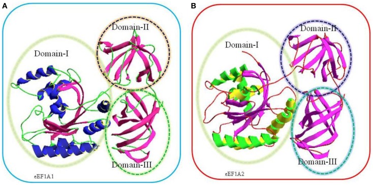
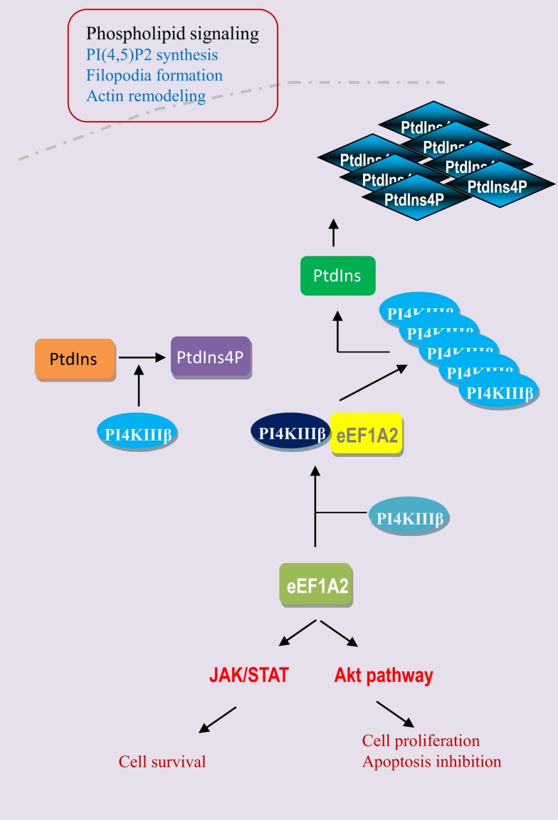
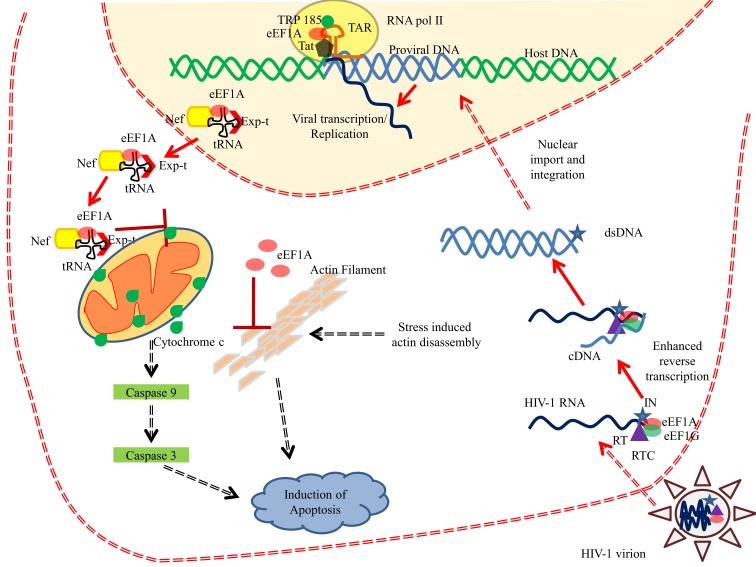
Eukaryotic translation elongation factors 1 alpha, eEF1A1 and eEF1A2, are not only translation factors but also pleiotropic proteins that are highly expressed in human tumors, including breast cancer, ovarian cancer, and lung cancer. eEF1A1 modulates cytoskeleton, exhibits chaperone-like activity and also controls cell proliferation and cell death. In contrast, eEF1A2 protein favors oncogenesis as shown by the fact that overexpression of eEF1A2 leads to cellular transformation and gives rise to tumors in nude mice. The eEF1A2 protein stimulates the phospholipid signaling and activates the Akt-dependent cell migration and actin remodeling that ultimately favors tumorigenesis. In contrast, inactivation of eEF1A proteins leads to immunodeficiency, neural and muscular defects, and favors apoptosis. Finally, eEF1A proteins interact with several viral proteins resulting in enhanced viral replication, decreased apoptosis, and increased cellular transformation. This review summarizes the recent findings on eEF1A proteins indicating that eEF1A proteins play a critical role in numerous human diseases through enhancement of oncogenesis, blockade of apoptosis, and increased viral pathogenesis.

Keywords: HIV; apoptosis; cancer; eEF1A; virus.


Figures

Figure 1
Figure 1
Comparative three-dimensional (3-D) model of eEF1A1 (A) and eEF1A2 (B) on the basis of crystal structure of homologous eEF1A from yeast. The target sequences used were eEF1A1 (Swiss-Prot Accession No: P68104) and eEF1A2 (Swiss-Prot Accession No: Q05639). The amino acids sequences of each protein were submitted to SWISS-MODEL server to build a 3D model (10).The highest resolved structure, 1.67-Å X-ray-derived eEF1A protein structure from yeast (Saccharomyces cerevisiae) with PDB ID: 1f60 and E-value 0.0 (sequence identity: 80.371%) was used as a template for modeling. Structurally, each model consists of three domains, domain I, domain II, and domain III as shown in the above cartoon. Domain I (residues 1–240) is made up of Rossmann-fold topology. Domain II (residues 241–336) and domain III (residues 337–443) are made up of entirely from beta-strands; each domain contains two beta sheets that form a beta barrel (7).
Figure 2
Figure 2
Phylogenetic tree constructed from the alignment of nucleotide and protein sequences of eEF1A2. The horizontal lines are the branches and suggest the amount of evolutionary changes over time. (A) Phylogenetic tree based on nucleotide sequences of eEF1A2 (Human: NM_001958.3, Chimpanzee: XM_003954094.1, Rat: NM_012660.2, Mouse: NM_007906.2, Fruit fly: NM_079872.4, Chicken: NM_001032398.2, and Mosquito: XM_003436467.1) using neighbor-joining distance method. The numbers indicate the evolutionary distances. (B) Phylogenetic tree based on amino acid sequences of eEF1A2 (Human: NP_001949.1, Chimpanzee: XP_003954143.1, Rat: NP_036792.2, Mouse: NP_031932.1, Fruit fly: NP_524611.1, Chicken: NP_001027570.1, and Mosquito: XP_003436515.1 using neighbor-joining (PAM250). The Jalview program was used for tree construction.
Figure 3
Figure 3
Promoter of eEF1A2. The promoter of eEF1A2 spans approximately 2.26 kb. Analysis of promoter sequence reveals 13 E-boxes, 3 EGR binding sites, and 1 MEF2 binding site. The core promoter region spans from position −16 to +92 bp.
Figure 4
Figure 4
eEF1A2 activates the phospholipid, JAK/STAT, and Akt pathways. eEF1A2 is a physiological regulator of phosphatidylinositol-4-kinase (PI4K). It directly interacts with phosphatidyloinositol-4 kinase III β (PI4KIIIβ) and enhances its lipid kinase activity by converting the phosphatidylinositol (PtdIns) into phosphatidylinositol-4-phosphate (PtdIns4P). Then PI4KIIIβ and PtdIns4P increase the phosphatidylinositol-4,5 bisphosphate generation at the plasma membrane level, which results in the filopodia formation and actin remodeling. eEF1A2 also directly or indirectly regulates the JAK/STAT and Akt signaling pathways.
Figure 5
Figure 5
eEF1A interferes with HIV-1 replication. eEF1A plays important role in various phases of HIV-1 life cycle. eEF1A has been reported in the mature HIV-1 virion and is also found to be a part of reverse transcription complex (RTC). Binding of eEF1A/EF1G to the RTC resulted in enhanced reverse transcription. In addition, eEF1A also help in the recruitment of RNA polymerase II and TRP-185 to the TAR RNA, which in turn regulates the viral transcription from 5′LTR. Moreover, HIV-1 Nef has been shown to interact with eEF1A and resulted in nucleo-cytoplasmic shuttling of eEF1A and ultimately in the inhibition of stress-induced apoptosis. Role of eEF1A in inhibiting the actin filament disassembly has been also proposed (77). Abbreviations: eEF1A, eukaryotic translation elongation factor 1 alpha; Exp-t, exportin-t; dsDNA, double stranded DNA; cDNA, complementary DNA; IN, integrase; RT, reverse transcriptase; RTC, reverse transcription complex; tRNA, transfer RNA; TAR, trans-activation response element; RNA pol II, RNA polymerase II; Tat, transactivator protein.

References

    1. Thornton S, Anand N, Purcell D, Lee J. Not just for housekeeping: protein initiation and elongation factors in cell growth and tumorigenesis. J Mol Med (Berl) (2003) 81(9):536–48.10.1007/s00109-003-0461-8 - DOI - PubMed
    1. Li GW, Xie XS. Central dogma at the single-molecule level in living cells. Nature (2011) 475(7356):308–15.10.1038/nature10315 - DOI - PMC - PubMed
    1. Garner AL, Janda KD. Protein-protein interactions and cancer: targeting the central dogma. Curr Top Med Chem (2011) 11(3):258–80.10.2174/156802611794072614 - DOI - PubMed
    1. Caraglia M, Budillon A, Vitale G, Lupoli G, Tagliaferri P, Abbruzzese A. Modulation of molecular mechanisms involved in protein synthesis machinery as a new tool for the control of cell proliferation. Eur J Biochem (2000) 267(13):3919–36.10.1046/j.1432-1327.2000.01465.x - DOI - PubMed
    1. Clemens MJ. Targets and mechanisms for the regulation of translation in malignant transformation. Oncogene (2004) 23(18):3180–8.10.1038/sj.onc.1207544 - DOI - PubMed