. 2015 Jan 29;Suppl 2(1):1-6.

Retinal Pigment Epithelial Cell Line Suppression of Phagolysosome Activation

Affiliations

Retinal Pigment Epithelial Cell Line Suppression of Phagolysosome Activation

A W Taylor et al. Int J Ophthalmol Eye Sci. .

Abstract

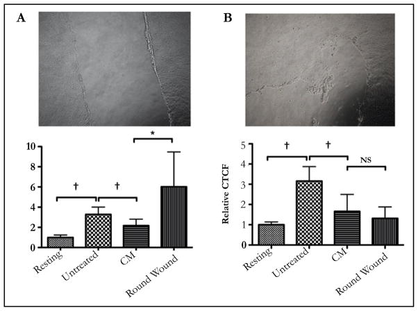
The eye is an immune privileged tissue with multiple mechanisms of immunosuppression to protect the light gathering tissues from the damage of inflammation. One of theses mechanisms involves retinal pigment epithelial cell suppression of phagosome activation in macrophages. The objective of this work is to determine if the human RPE cell line ARPE-19 is capable of suppressing the activation of the phagolysosome in macrophages in a manner similar to primary RPE. The conditioned media of RPE eyecups, sub-confluent, just confluent cultures, or established confluent cultures of human ARPE-19 cells were generated. These condition media were used to treat macrophages phagocytizing pHrodo bioparticles. After 24 hours incubation the macrophages were imaged by fluorescent microscopy, and fluorescence was measured. The fluorescent intensity is proportional to the amount of bioparticles phagocytized and are in an activated phagolysosome. The conditioned media of in situ mouse RPE eyecups significantly suppressed the activation of phagolysosome. The conditioned media from cultures of human ARPE-19 cells, grown to sub-confluence (50%) or grown to confluence had no effect on phagolysosome activation. In contrast, the conditioned media from established confluent cultures significantly suppressed phagolysosome activation. The neuropeptides alpha-MSH and NPY were depleted from the conditioned media of established confluent ARPE-19 cell cultures. This depleted conditioned media had diminished suppression of phagolysosome activation while promoting macrophage cell death. In addition, the condition media from cultures of ARPE-19 monolayers wounded with a bisecting scrape was diminished in suppressing phagolysosome activation. This technical report suggests that like primary RPE monolayers, established confluent cultures of ARPE-19 cells produce soluble factors that suppress the activation of macrophages, and can be used to study the molecular mechanisms of retinal immunobiology. In addition, the results further demonstrate the importance of an intact monolayer of RPE cells to modulate immune cell activity within the eye.

Keywords: Alpha-Melanocyte Stimulating Hormone; Immune Privilege; Macrophages; Neuroimmunomodulation; Neuropeptide Y; Phagocytosis.


Figures

Figure 1
Figure 1. The conditioned media of RPE eyecups suppresses pHrodo-bioparticle fluorescence in macrophages. The macrophages were cultured as described, and treated with the conditioned media of RPE eye cups (CM) for 24 hours
A) Representative images of the florescence observed with resting, untreated, and CM-treated macrophages. B) The corrected total cellular fluorescence (CTCF) was calculated and made relative to macrophages that were not given beads (Resting). The results are the mean ± SD of 4 cultures analyzing a total of 80 – 100 cells per condition. *Significantly different P ≤ 0.01, and NS is not significant. †P≤ 0.001. The RPE conditioned media suppressed the fluorescence of pHrodo-bioparticles in macrophages indicating suppression of the activation of the phagolysosome.
Figure 2
Figure 2. The conditioned media from established ARPE-19 confluent cultures suppressed pHrodo-bioparticle fluorescence in macrophages
A) The ARPE-19 cells were grown to confluence, or kept subconfluent before changing the media for serum-free media. After 24 hours incubation in serum-free media the conditioned media (CM) was collected, and the macrophages were treated with the CM before fed pHrodo-bioparticles. 24 hours later the cells were assayed for fluorescence. No significant difference was seen between the pHrodo-bioparticle fluorescence in macrophages treated with CM from confluent or subconfluent cultures with untreated macrophages. B)The confluent ARPE-19 cultures were incubated for an additional 2 days before the media was changed to serum-free. *Significant (P≤0.01) differences were seen between the established confluent CM treated macrophages and the untreated macrophages. NS is not significant. †P ≤ 0.01. The results are the mean ± SD of 4 cultures analyzing a total of 50 – 75 cells per condition. Only the established confluence cultures of ARPE-19 cell produced soluble factors that suppressed the activation of the phagolysosome.
Figure 3
Figure 3. The depletion of α-MSH and NPY from CM of established confluent APRE-19 cultures removes the suppression of pHrodo-bioparticle fluorescence in macrophages
A) The conditioned media of established ARPE-19 cell cultures antibody depleted of α-MSH and NPY were used to treat the macrophages that were then fed pHrodo-bioparticles. *There was a significant (P ≤ 0.01) increase in the pHrodo-bioparticles fluorescence in macrophages treated with CM depleted of α-MSH and NPY (CM-MSH-NPY) with macrophages treated with CM absorbed with irrelevant antibody (CM(IgGAb)). This was not significantly different from the untreated macrophages. Presented are the mean ± SD of 4 cultures analyzing a total of 50 – 75 cells per condition. B) The images of macrophages treated with CM(IgGAb) was compared with the images of the macrophages treated with the α-MSH/NPY-depleted CM. Arrows indicate dead or possible apoptotic cells. The arrow with the star marks the cell that was magnified in the figure inset. The ARPE-19 suppression of phagolysosome activation is mediated by at least α-MSH and NPY, and their depletion from the conditioned media causes macrophage cell death similar to what has been reported for conditioned media of RPE eyecups [4].
Figure 4
Figure 4. The effects of disrupting the ARPE-19 established confluent monolayer. Established ARPE-19 confluent monolayers were
A) bisected with a scrape, or B) wounded with round scrapes before the cell were cultured in serum-free media. Presented are representative phase contract micrographs of the wounded cultures and the relative effects of the conditioned media on phagolysosome activation. *Significant (P ≤ 0.05) loss of suppression was seen with the conditioned media from ARPE-19 cultures with a bisecting scrape wound, but not with rounded wounds. Moreover the pHrodo-bioparticles fluorescence in macrophages treated with CM from the bisected scrape cultures had enhanced levels of fluorescence. †P ≤ 0.05. The results are the mean ± SD of 4 cultures analyzing a total of 50 – 75 cells per condition. An intact confluent monolayer necessary for ARPE-19 cells to suppress the activation of the phagolysosome.

References

    1. Zamiri P, Masli S, Kitaichi N, Taylor A, Streilein J. Thrombospondin plays a vital role in the immune privilege of the eye. Investigative Ophthalmology & Visual Science. 2005;46:908–919. - PubMed
    1. Zamiri P, Masli S, Streilein JW, Taylor AW. Pigment epithelial growth factor suppresses inflammation by modulating macrophage activation. Investigative Ophthalmology & Visual Science. 2006;47:3912–3918. - PubMed
    1. Lau CH, Taylor AW. The immune privileged retina mediates an alternative activation of J774A.1 cells. Ocul Immunol Inflamm. 2009;17:380–9. - PMC - PubMed
    1. Kawanaka N, Taylor AW. Localized retinal neuropeptide regulation of macrophage and microglial cell functionality. Journal of Neuroimmunology. 2011;232:17–25. - PMC - PubMed
    1. Tu Z, Li Y, Smith D, Doller C, Sugita S, et al. Myeloid suppressor cells induced by retinal pigment epithelial cells inhibit autoreactive T-cell responses that lead to experimental autoimmune uveitis. Investigative ophthalmology & visual science. 2012;53:959–66. - PMC - PubMed

LinkOut - more resources