. 2015 Dec;53(6):782-92.
doi: 10.1165/rcmb.2014-0474OC.

Disruption of Sirtuin 1-Mediated Control of Circadian Molecular Clock and Inflammation in Chronic Obstructive Pulmonary Disease

Affiliations

Disruption of Sirtuin 1-Mediated Control of Circadian Molecular Clock and Inflammation in Chronic Obstructive Pulmonary Disease

Hongwei Yao et al. Am J Respir Cell Mol Biol. 2015 Dec.

Abstract

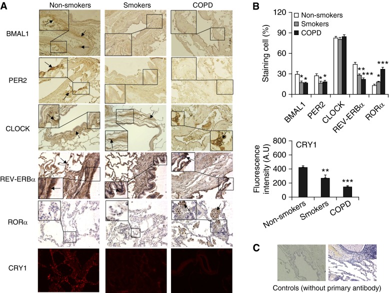
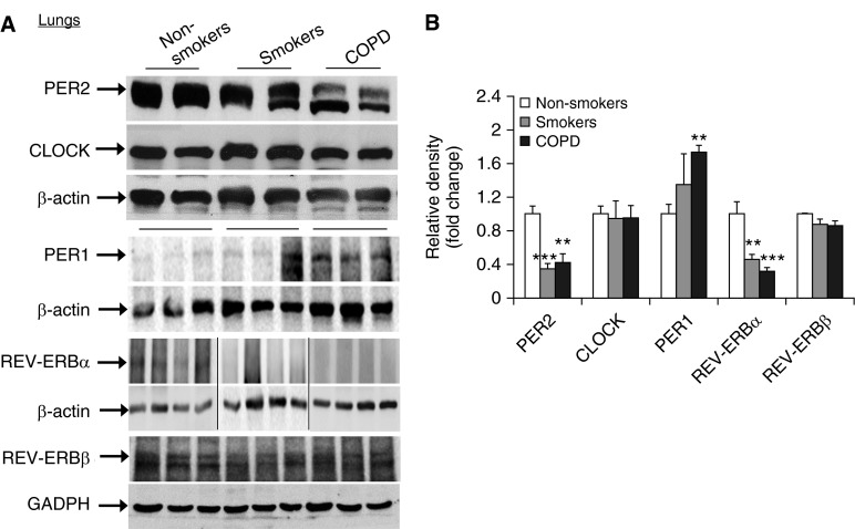
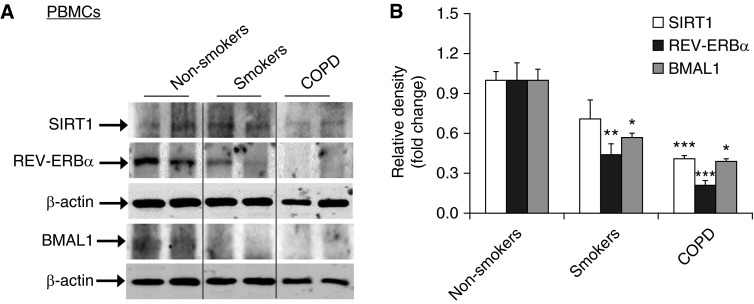
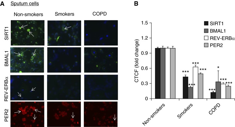
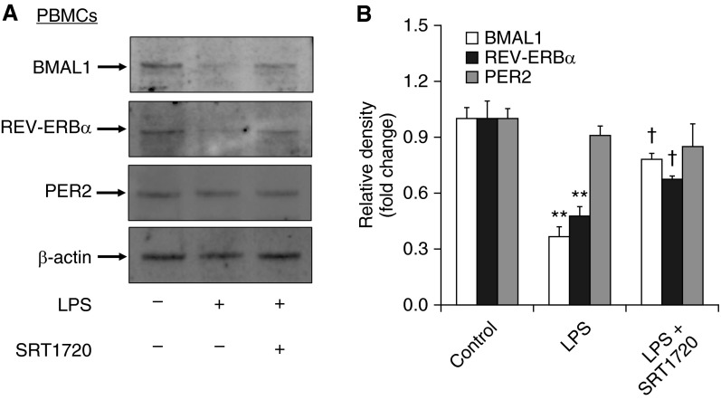
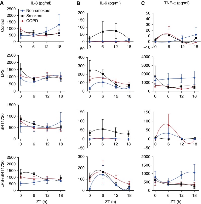
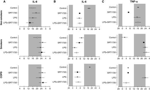
Chronic obstructive pulmonary disease (COPD) is the fourth most common cause of death, and it is characterized by abnormal inflammation and lung function decline. Although the circadian molecular clock regulates inflammatory responses, there is no information available regarding the impact of COPD on lung molecular clock function and its regulation by sirtuin 1 (SIRT1). We hypothesize that the molecular clock in the lungs is disrupted, leading to increased inflammatory responses in smokers and patients with COPD and its regulation by SIRT1. Lung tissues, peripheral blood mononuclear cells (PBMCs), and sputum cells were obtained from nonsmokers, smokers, and patients with COPD for measurement of core molecular clock proteins (BMAL1, CLOCK, PER1, PER2, and CRY1), clock-associated nuclear receptors (REV-ERBα, REV-ERBβ, and RORα), and SIRT1 by immunohistochemistry, immunofluorescence, and immunoblot. PBMCs were treated with the SIRT1 activator SRT1720 followed by LPS treatment, and supernatant was collected at 6-hour intervals. Levels of IL-8, IL-6, and TNF-α released from PBMCs were determined by ELISA. Expression of BMAL1, PER2, CRY1, and REV-ERBα was reduced in PBMCs, sputum cells, and lung tissues from smokers and patients with COPD when compared with nonsmokers. SRT1720 treatment attenuated LPS-mediated reduction of BMAL1 and REV-ERBα in PBMCs from nonsmokers. Additionally, LPS differentially affected the timing and amplitude of cytokine (IL-8, IL-6, and TNF-α) release from PBMCs in nonsmokers, smokers, and patients with COPD. Moreover, SRT1720 was able to inhibit LPS-induced cytokine release from cultured PBMCs. In conclusion, disruption of the molecular clock due to SIRT1 reduction contributes to abnormal inflammatory response in smokers and patients with COPD.

Keywords: BMAL1; REV-ERBα; SIRT1; circadian rhythm; smokers.


Figures

Figure 1.
Figure 1.
Changes of clock proteins and nuclear receptors in lungs from smokers and patients with chronic obstructive pulmonary disease (COPD) by immunostaining. (A) Expression of BMAL1, CLOCK, PER2, REV-ERBα, and RORα were measured by immunohistochemistry, and CRY1 was measured by immunofluorescence staining in lungs from nonsmokers, smokers, and patients with COPD. Original magnification: ×200. Dark brown color represents the presence of BMAL1, CLOCK, PER2, and REV-ERBα, and RORα, which are indicated with arrows. CRY1 presence is shown as red. The insets show high magnification of clock protein–positive cells. (B) Immunostaining scores for BMAL1, CLOCK, PER2, REV-ERBα, and RORα and fluorescence intensity with arbitrary units (A.U.) for CRY1 in lung cells were performed semiquantitatively and in a blinded fashion. Data are shown as mean ± SEM (n = 4–6). *P < 0.05, **P < 0.01, and ***P < 0.001 versus nonsmokers. (C) Representative images of lung tissue stained without primary antibody as a control.
Figure 2.
Figure 2.
Levels of PER2, CLOCK, PER1, REV-ERBα, and REV-ERBβ in lungs from nonsmokers, smokers, and patients with COPD by Western blot. (A) Levels of PER2, CLOCK, PER1, REV-ERBα, and REV-ERBβ in lung tissues from nonsmokers, smokers, and patients with COPD were measured by Western blot. Each group represents the results of at least three independent experiments, and the representative REV-ERBα bands are from different gels, which are demarcated by the lines. (B) The relative band intensity normalized to β-actin or glyceraldehyde 3-phosphate dehydrogenase (GAPDH) is shown in a histogram. Data are shown as mean ± SEM (n = 3–4). **P < 0.01 and ***P < 0.001 versus nonsmokers.
Figure 3.
Figure 3.
Reduction of clock proteins along with sirtuin 1 (SIRT1) levels in peripheral blood mononuclear cells (PBMCs) from smokers and patients with COPD. (A) Levels of REV-ERBα and BMAL1 as well as SIRT1 in PBMCs from nonsmokers, smokers, and patients with COPD were measured by Western blot. Each group represents the results of five independent experiments, and the representative bands, which are demarcated by the lines based on groups, are from the same gel. (B) The relative band intensity normalized to GAPDH or β-actin is shown in the histogram. Data are shown as mean ± SEM (n = 3–4). *P < 0.05, **P < 0.01, and ***P < 0.001 versus nonsmokers.
Figure 4.
Figure 4.
Expression of clock proteins along with SIRT1 levels in sputum cells from nonsmokers, smokers, and patients with COPD. (A) The abundance of clock machinery proteins REV-ERBα, BMAL1, and PER2 was measured by immunofluorescent staining in sputum cells from nonsmokers, smokers, and patients with COPD. SIRT1 and BMAL1 are shown in green, 4′,6-diamidino-2-phenylindole is shown in blue, and PER2 expression is shown in red. Results are representative cells of at least three separate experiments. The arrows indicate the positive cells of SIRT1, BMAL1, REV-ERBα, and PER2. (B) The quantification of fluorescence intensity in immunofluorescence data was measured using ImageJ, and the corrected total cell fluorescence (CTCF) values were converted into fold change values and represented as histograms. Data are shown as mean ± SEM (n = 5). *P < 0.05 and ***P < 0.001 versus nonsmokers.
Figure 5.
Figure 5.
SRT1720 treatment attenuated LPS-induced reduction of REV-ERBα and BMAL1 in PBMCs. (A) The levels of REV-ERBα, BMAL1, and PER2 were measured by Western blot in LPS-treated PBMCs from nonsmokers. Each group represents the results of at least three independent experiments. (B) The relative band intensity normalized to β-actin is shown in the histogram. Data are shown as mean ± SEM (n = 3). **P < 0.01 versus controls; P < 0.05 versus LPS treatment.
Figure 6.
Figure 6.
Circadian rhythms of proinflammatory mediator release in LPS-treated PBMCs from nonsmokers, smokers, and patients with COPD and their regulation by SRT1720. PBMCs from nonsmokers, smokers, and patients with COPD were treated with SRT1720 (1 μM) for 24 hours followed by 2 hours of LPS (1 μg/ml) treatment before supernatant collection at each 6-hour interval. Levels of IL-8 (A), IL-6 (B), and TNF-α (C) in culture supernatants were determined by ELISA. Data were fit with nonlinear regression (multiorder polynomial) analyses. Data are shown as mean ± SEM (n = 6–9 per group). ZT, Zeitgeber time.
Figure 7.
Figure 7.
Phase values of proinflammatory mediator release in LPS-treated PBMCs and its regulation by SRT1720. Center of gravity or peak phase values for IL-8 (A), IL-6 (B), and TNF-α (C) expression rhythm in PBMCs from nonsmokers, smokers, and patients with COPD were obtained by using CircWave and plotted on a horizontal phase map. Gray shading indicates the relative dark phase (ZT12–ZT24). Data are shown as mean ± SEM (n = 6–9 per group).

Similar articles

Cited by

References

    1. Tsai CL, Brenner BE, Camargo CA., Jr Circadian-rhythm differences among emergency department patients with chronic obstructive pulmonary disease exacerbation. Chronobiol Int. 2007;24:699–713. - PubMed
    1. Borsboom GJ, van Pelt W, van Houwelingen HC, van Vianen BG, Schouten JP, Quanjer PH. Diurnal variation in lung function in subgroups from two Dutch populations: consequences for longitudinal analysis. Am J Respir Crit Care Med. 1999;159:1163–1171. - PubMed
    1. Hwang JW, Sundar IK, Yao H, Sellix MT, Rahman I. Circadian clock function is disrupted by environmental tobacco/cigarette smoke, leading to lung inflammation and injury via a SIRT1-BMAL1 pathway. FASEB J. 2014;28:176–194. - PMC - PubMed
    1. Hoegh SV, Sorensen GL, Tornoe I, Lottenburger T, Ytting H, Nielsen HJ, Junker P, Holmskov U. Long-term stability and circadian variation in circulating levels of surfactant protein D. Immunobiology. 2010;215:314–320. - PubMed
    1. Gibbs J, Ince L, Matthews L, Mei J, Bell T, Yang N, Saer B, Begley N, Poolman T, Pariollaud M, et al. An epithelial circadian clock controls pulmonary inflammation and glucocorticoid action. Nat Med. 2014;20:919–926. - PMC - PubMed

Publication types