. 2015 Apr 23;10(4):e0124227.
doi: 10.1371/journal.pone.0124227. eCollection 2015.

Safety of intracoronary infusion of 20 million C-kit positive human cardiac stem cells in pigs

Affiliations

Safety of intracoronary infusion of 20 million C-kit positive human cardiac stem cells in pigs

Matthew C L Keith et al. PLoS One. .

Abstract

Background: There is mounting interest in using c-kit positive human cardiac stem cells (c-kit(pos) hCSCs) to repair infarcted myocardium in patients with ischemic cardiomyopathy. A recent phase I clinical trial (SCIPIO) has shown that intracoronary infusion of 1 million hCSCs is safe. Higher doses of CSCs may provide superior reparative ability; however, it is unknown if doses >1 million cells are safe. To address this issue, we examined the effects of 20 million hCSCs in pigs.

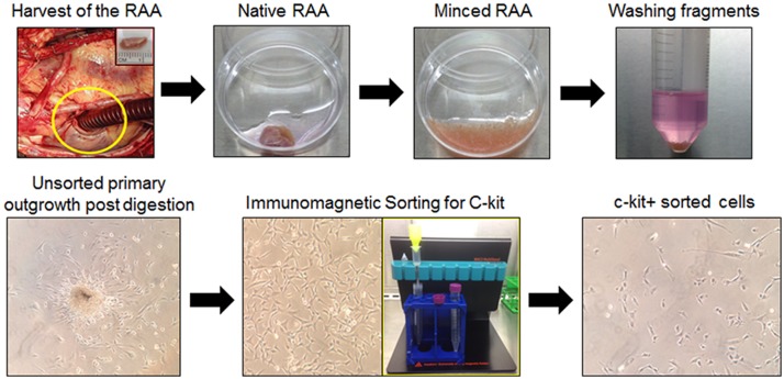
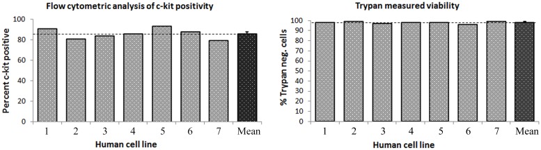
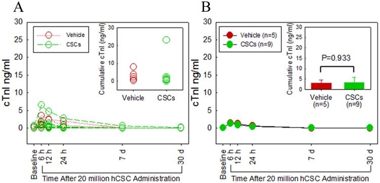
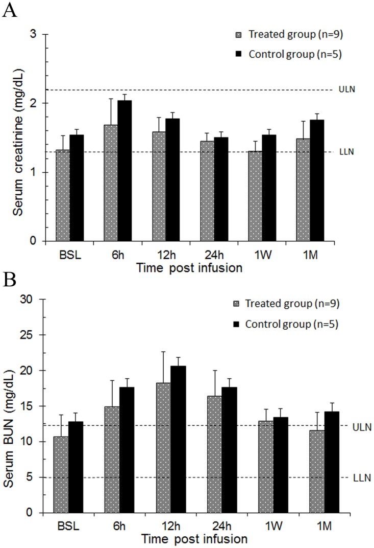
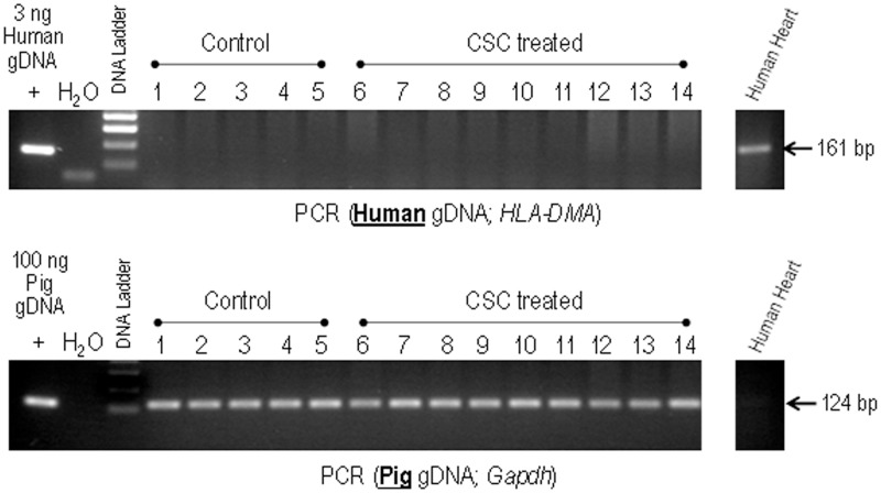
Methods: Right atrial appendage samples were obtained from patients undergoing cardiac surgery. The tissue was processed by an established protocol with eventual immunomagnetic sorting to obtain in vitro expanded hCSCs. A cumulative dose of 20 million cells was given intracoronarily to pigs without stop flow. Safety was assessed by measurement of serial biomarkers (cardiac: troponin I and CK-MB, renal: creatinine and BUN, and hepatic: AST, ALT, and alkaline phosphatase) and echocardiography pre- and post-infusion. hCSC retention 30 days after infusion was quantified by PCR for human genomic DNA. All personnel were blinded as to group assignment.

Results: Compared with vehicle-treated controls (n=5), pigs that received 20 million hCSCs (n=9) showed no significant change in cardiac function or end organ damage (assessed by organ specific biomarkers) that could be attributed to hCSCs (P>0.05 in all cases). No hCSCs could be detected in left ventricular samples 30 days after infusion.

Conclusions: Intracoronary infusion of 20 million c-kit positive hCSCs in pigs (equivalent to ~40 million hCSCs in humans) does not cause acute cardiac injury, impairment of cardiac function, or liver and renal injury. These results have immediate translational value and lay the groundwork for using doses of CSCs >1 million in future clinical trials. Further studies are needed to ascertain whether administration of >1 million hCSCs is associated with greater efficacy in patients with ischemic cardiomyopathy.


Conflict of interest statement

Competing Interests: The authors have declared that no competing interests exist.

Figures

Fig 1
Fig 1. Study protocol and timeline.
Fig 2
Fig 2. Isolation and expansion of c-kitpos hCSCs.
Right atrial appendages (RAA) were harvested with subsequent mechanical and enzymatic digestion to obtain primary outgrowth of total adherent cardiac cells. Primary cells were immunomagnetically sorted for c-kit and the resultant cells expanded in vitro.
Fig 3
Fig 3. Flow cytometric validation and immunocytochemistry of c-kitpos hCSCs.
Representative flow cytometric analyses of isotype control (left) and c-kit-labeled cell flow plots (center) are shown. Suspension immunocytochemistry of c-kitpos hCSCs showing positive anti-c-kit labeling is shown in the right panels, with DAPI labeled nuclei in blue.
Fig 4
Fig 4. Cumulative c-kit positivity by flow cytometry and Trypan blue cell product viability.
The left panel shows c-kit positivity in seven cell lines utilized for the study, which averaged 85.6%±1.9% (mean±SEM). The right panel shows viability of c-kitpos hCSCs measured by cellular exclusion of Trypan blue staining prior to intracoronary infusion. Trypan negative, viable cells averaged 97.8±0.4%. Data are mean±SEM.
Fig 5
Fig 5. Intracoronary infusion of 20 million human c-kitpos CSCs does not impair left ventricular (LV) function or morphology.
The line graphs in the top panel show individual values of each pig’s progress over time (baseline, 6, 12, 24 h, 1 week, and 1 month). Individual plots are in yellow, and group means are illustrated by the red line plots. A. ejection fraction (EF). B. LV end-diastolic diameter (EDD). C. LV end-diastolic volume (EDV), D. LV anterior wall fractional thickening. The bottom panel shows group mean±SEM at each time point for respective LV functional and morphologic indices. Green and red bars indicate hCSC-treated and vehicle groups respectively. E. ejection fraction, F. fractional shortening, G. LV end-systolic area, H. LV end-diastolic area, I. LV end-systolic volume, J. LV end-diastolic volume, K. LV anterior wall thickness in systole, L. LV anterior wall thickness in diastole, M. LV end-systolic diameter, N. LV end-diastolic diameter, O. LV posterior wall thickness in systole, P. LV posterior wall thickness in diastole. Data are mean±SEM. There were no significant differences between groups with respect to any parameter at respective time points (P > 0.05).
Fig 6
Fig 6. Intracoronary infusion of 20 million human c-kitpos CSCs does not cause myocardial damage as assessed by cardiac troponin I (cTnI) release.
A. Individual plots of serum cTnI levels (ng/ml) are shown at serial time points (baseline, 6, 12, 24 h, 1 week, and 1 month). Green and red plots indicate hCSC-treated and vehicle control pigs, respectively. B. Group means at each time point are shown. The green and red plots indicate hCSC-treated and vehicle control groups, respectively. The inset in panel B shows cumulative cTnI levels. Data are mean±SEM. There were no significant differences in plasma cTnI levels over the 1 month follow up between groups (P > 0.05 at each time point).
Fig 7
Fig 7. Intracoronary infusion of 20 million human c-kitpos CSCs does not cause myocardial damage as assessed by cardiac CK-MB release.
A. Individual serum CK-MB levels (ng/ml) over serial time points (baseline, 6, 12, 24 h, 1 week, and 1 month). Green and red plots identify hCSC-treated and vehicle control pigs, respectively. B. Group means at each time point are shown. The green and red plots identify hCSC-treated and vehicle control groups, respectively. The inset in panel B shows cumulative CK-MB levels. Data are mean±SEM. There were no significant differences in plasma CK-MB levels over the 1 month follow up between groups (P > 0.05 at each time point).
Fig 8
Fig 8. Intracoronary infusion of 20 million human c-kitpos CSCs does not impair renal function.
Renal function was assessed by serum creatinine and blood urea nitrogen (BUN) values over serial time-points (baseline, 6, 12, 24 h, 1 week, and 1 month). A. Bar graph of serum creatinine levels in hCSC-treated and vehicle control group. B. Bar graph of serum BUN levels in hCSC-treated and vehicle control group. Blue and purple bars identify hCSC-treated and vehicle control groups, respectively. Upper limits of normal (ULN) and lower limits of normal (LLN) in each graph are depicted by dashed lines respectively. Data are mean±SEM. There were no significant differences in serum creatinine or BUN levels over the 1 month follow up between groups (P > 0.05 at each time point).
Fig 9
Fig 9. Intracoronary infusion of 20 million human c-kitpos CSCs does not impair liver function.
Liver function was assessed by serum AST, ALT, alkaline phosphatase, and total CK levels at serial time points (baseline, 6, 12, 24 h, 1 week, and 1 month). A. Serum aspartate aminotransferase (AST), B. Serum creatine phosphokinase (CPK), C. Serum alkaline phosphatase (Alk. Phos.), D. Serum alanine aminotransferase (ALT). Upper limits of normal) and lower limits of normal (LLN) in each graph are depicted by dashed lines respectively. Data are mean±SEM. There were no significant differences in serum AST, ALT, alkaline phosphatase, or total CK levels over the 1 month follow up between groups (P > 0.05 at each time point).
Fig 10
Fig 10. Detection of human CSCs in control versus hCSC-treated pig hearts.
Genomic DNA isolated from representative LV sections from control (lanes 1–5) and human CSC-treated pigs (lanes 6–14) were analyzed by PCR for the presence of human genomic DNA (HLA-DMA). Samples were also analyzed for the presence of pig genomic DNA (Gapdh) as a control for DNA quality. Genomic DNA isolated from human heart sections was used as both positive and negative control. None of the samples, including CSC-treated ones, show detectable levels of human DNA.

References

    1. Beltrami AP, Barlucchi L, Torella D, Baker M, Limana F, Chimenti S, et al. Adult cardiac stem cells are multipotent and support myocardial regeneration. Cell. 2003;114: 763–776. - PubMed
    1. Bolli R, Chugh AR, D'Amario D, Loughran JH, Stoddard MF, Ikram S, et al. Cardiac stem cells in patients with ischaemic cardiomyopathy (SCIPIO): initial results of a randomised phase 1 trial. Lancet. 2011;378: 1847–1857. 10.1016/S0140-6736(11)61590-0 - DOI - PMC - PubMed
    1. Meluzin J, Mayer J, Groch L, Janousek S, Hornacek I, Hlinomaz O, et al. Autologous transplantation of mononuclear bone marrow cells in patients with acute myocardial infarction: the effect of the dose of transplanted cells on myocardial function. Am Heart J. 2006;152: 975 e979–915. - PubMed
    1. Katritsis DG, Sotiropoulou PA, Karvouni E, Karabinos I, Korovesis S, Perez SA, et al. Transcoronary transplantation of autologous mesenchymal stem cells and endothelial progenitors into infarcted human myocardium. Catheter Cardiovasc Interv. 2005;65: 321–329. - PubMed
    1. Gao LR, Pei XT, Ding QA, Chen Y, Zhang NK, Chen HY, et al. A critical challenge: dosage-related efficacy and acute complication intracoronary injection of autologous bone marrow mesenchymal stem cells in acute myocardial infarction. Int J Cardiol. 2013;168: 3191–3199. 10.1016/j.ijcard.2013.04.112 - DOI - PubMed

Publication types

Substances