. 2015 Apr 3;11(4):670-84.
doi: 10.1080/15548627.2015.1034409.

Essential role for the ATG4B protease and autophagy in bleomycin-induced pulmonary fibrosis

Affiliations

Essential role for the ATG4B protease and autophagy in bleomycin-induced pulmonary fibrosis

Sandra Cabrera et al. Autophagy. .

Abstract

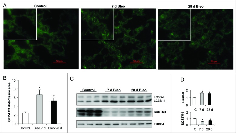
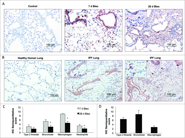
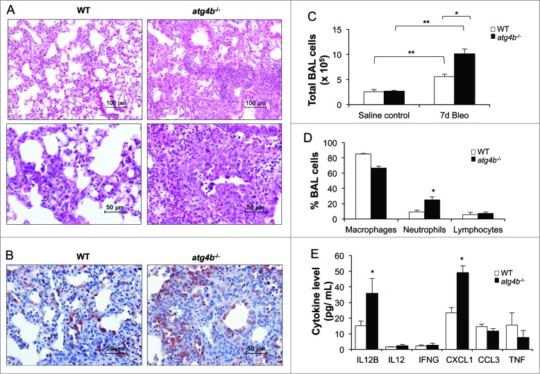
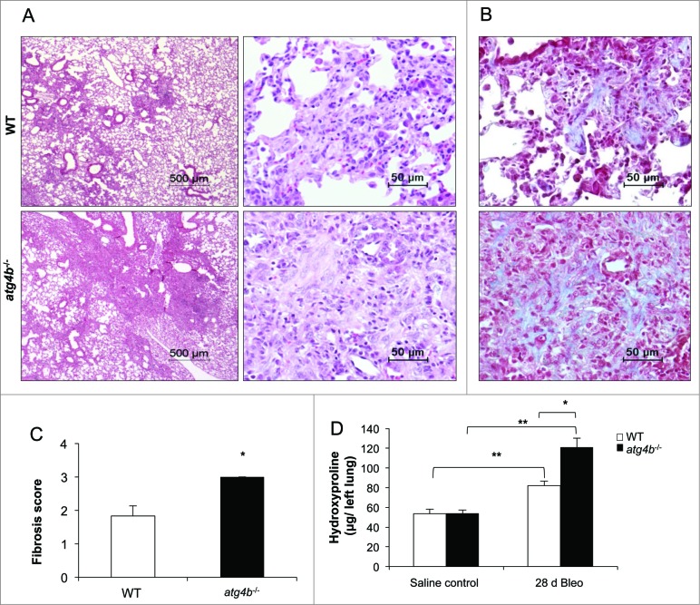
Autophagy is a critical cellular homeostatic process that controls the turnover of damaged organelles and proteins. Impaired autophagic activity is involved in a number of diseases, including idiopathic pulmonary fibrosis suggesting that altered autophagy may contribute to fibrogenesis. However, the specific role of autophagy in lung fibrosis is still undefined. In this study, we show for the first time, how autophagy disruption contributes to bleomycin-induced lung fibrosis in vivo using an Atg4b-deficient mouse as a model. Atg4b-deficient mice displayed a significantly higher inflammatory response at 7 d after bleomycin treatment associated with increased neutrophilic infiltration and significant alterations in proinflammatory cytokines. Likewise, we found that Atg4b disruption resulted in augmented apoptosis affecting predominantly alveolar and bronchiolar epithelial cells. At 28 d post-bleomycin instillation Atg4b-deficient mice exhibited more extensive and severe fibrosis with increased collagen accumulation and deregulated extracellular matrix-related gene expression. Together, our findings indicate that the ATG4B protease and autophagy play a crucial role protecting epithelial cells against bleomycin-induced stress and apoptosis, and in the regulation of the inflammatory and fibrotic responses.

Keywords: ACTA2, actin, α 2, smooth muscle, aorta; ATG3, autophagy-related 3; ATG4B; ATG4B, autophagy-related 4B; ATG5, autophagy-related 5; ATG7, autophagy-related 7; ATG9B, autophagy-related 9B; BAX, BCL2-associated X protein; CASP3, caspase 3, apoptosis-related cysteine peptidase; CAV1, caveolin 1, caveolae protein, 22kDa; CCL3, chemokine (C-C motif) ligand 3; CXCL1, chemokine (C-X-C motif) ligand 1 (melanoma growth stimulating activity α); CXCR2, chemokine (C-X-C motif) receptor 2; DRAM2, DNA-damage regulated autophagy modulator 2; GFP-LC3B, green fluorescent protein-LC3B; IFNG, interferon, gamma; IL12B, interleukin 12B; IL13, interleukin 13; IPF, idiopathic pulmonary fibrosis; MAP1LC3B/LC3B, microtubule-associated protein 1 light chain 3 β; RELA, v-rel reticuloendotheliosis viral oncogene homolog A; SQSTM1, sequestosome 1; TGFB1, transforming growth factor, β 1; TGFBR2, transforming growth factor, β receptor II (70/80kDa); TNF, tumor necrosis factor; TUBB4, tubulin, β 4, class IV; WT, wild type; autophagin-1; autophagy; cysteine peptidase; epithelial cell; idiopathic pulmonary fibrosis; lung fibrosis.


Figures

Figure 1.
Figure 1.
Autophagic activity increases after lung inflammation and fibrosis. Bleomycin (Bleo) or saline were administered to GFP-LC3 transgenic mice by endotracheal instillation and lungs were harvested after indicated time points. (A) Representative fluorescent microphotographs of lung tissue from saline control and bleomycin-treated GFP-LC3 transgenic mice. (B) Quantification of LC3B dots per mm2 of lung tissue from GFP-LC3 transgenic mice. (C) Representative immunoblots of endogenous LC3B-I/II and SQSTM1 in lung tissue. TUBB4 was used as a loading control. (D) Densitometry. Protein levels were normalized to saline control. Results represent mean ± SD. Statistical significance was determined by one-way ANOVA (*P < 0.05).
Figure 2.
Figure 2.
Autophagic activity in MLE 12 cells increases after bleomycin challenge. (A) Representative fluorescent microphotographs of endogenous LC3B dots in MLE 12 cells under basal or bleomycin-treated condition, and LC3B dots per cell area (μm2 × 100). (B) Representative immunoblots of endogenous LC3B-I/-II and SQSTM1 in MLE 12 cells after 0 or 100 mU/ml of bleomycin alone, or combined with 10 μM chloroquine or 10 μM leupeptin for 24 h (left), and densitometry normalized to the basal control condition (right). TUBB4 was used as loading control. (C) LC3B-II and SQSTM1 ratio (protein level in the presence of inhibitor vs protein level in the absence of inhibitor) in bleomycin-treated compared to control cells. Results represent mean ± SD. Statistical significance was determined by one-way ANOVA (*P < 0.05, ** P < 0.01).
Figure 3.
Figure 3.
Gene expression changes after bleomycin-induced lung fibrosis. (A) Gene expression infogram for differentially expressed genes at 28 d after bleomycin exposure performed by qPCR. Every row represents a gene and every column a mouse lung sample (control vs fibrosis, n = 3 for each group). (B) Representative immunoblots of ATG3, ATG4B, ATG5, and ATG7 in lung tissue extracts from control and bleomycin-treated mice. TUBB4 was used as loading control. (C) Densitometry. Protein levels were normalized to saline control. Results represent mean ± SD. Statistical significance was determined by one-way ANOVA (*P < 0.05).
Figure 4.
Figure 4.
ATG4B immunolocalization in bleomycin-induced pulmonary fibrosis and IPF lung. (A) Representative photomicrographs of immunohistochemical staining for ATG4B in saline control and bleomycin-treated mouse lung at 7 and 28 d. (B) Healthy and IPF human lung tissue sections. Positive staining was observed in red. All sections were counterstained with hematoxylin. (C) Semiquantitative score of mouse and (D) human lung tissue sections. Results represent mean ± SD. Statistical significance was determined by one-way ANOVA (*P < 0.05).
Figure 5.
Figure 5.
Reduced autophagic flux in lung tissue from atg4b−/− mice. Representative immunoblots of ATG4B, LC3B and SQSTM1 in lung tissue extracts from (A) control (B) 7 and (C) 28 d after bleomycin treatment from age-matched WT and mutant mice. Lung tissue extracts from 3 mice per genotype are shown. TUBB4 was used as loading control. (D) Representative light microscopy images of H&E stained lung tissue sections from WT and atg4b−/− control mice. (E) Percentage of body weight changes over a 28-day period for atg4b−/− and WT mice after bleomycin instillation. (F) Percentage of survival were plotted for atg4b−/− (n = 18) and WT (n = 18) mice for 28 d after challenge with bleomycin. Statistical significance was determined by log-rank test or Student t test (*P < 0.05).
Figure 6.
Figure 6.
Increased lung inflammatory response in atg4b−/− mice. (A) Representative light microscopy images of H&E stained lung tissue sections from WT and atg4b−/− mice at 7 d after bleomycin instillation. (B) Immunohistochemical staining performed with specific antibody against RELA/NFκB p65 in lung tissue sections from WT and atg4b−/− mice at 7 d post-bleomycin. (C) Total and (D) differential cell count in BALF. (E) Protein level of proinflammatory cytokines in BALF from atg4b−/− and WT bleomycin-treated mice. Results are shown as mean ± SD. Statistical significance was determined by one-way ANOVA (C) or Student t test (D and E) (*P < 0.05).
Figure 7.
Figure 7.
Atg4b deficiency sensitizes mice to experimental lung fibrosis. (A) Representative light microscopy images of H&E stained lung tissue sections from WT and atg4b−/− mice at 28 d after bleomycin instillation. (B) Masson Trichrome staining of lung sections from the same experimental groups. (C) Fibrosis score for grading lung histopathological changes. (D) Hydroxyproline content in lung at 28 d after bleomycin. Results are shown as mean ±SD . Statistical significance was determined by Student t test (C) or one-way ANOVA (D) (*P < 0.05).
Figure 8.
Figure 8.
Deregulated expression of profibrotic mediators in atg4b−/− mice. (A) Representative light microscopy images of stained lung tissue sections performed with specific antibody against ACTA2 from WT and atg4b−/− mice at 28 d after bleomycin instillation. (B) Representative immunoblot of ACTA2 in lung tissue extracts from WT and atg4b−/− mice at 28 d after bleomycin instillation and densitometry analysis. TUBB4 was used as loading control. Results are shown as mean ± SD. Statistical significance was determined by Student t test (*P < 0.05). (C) Gene expression infogram for differentially expressed genes at 28 d after bleomycin instillation performed by qPCR. Every row represents a gene and every column a mouse lung sample (WT vs atg4b−/− mice, n = 3 for each group).
Figure 9.
Figure 9.
Mice with Atg4b deficiency display increased apoptosis after bleomycin challenge. (A) Lung tissue sections labeled with TUNEL assay and counterstained with methyl green. Apoptotic DAB-positive nuclei are brown, normal nuclei are green. (B) Representative immunoblots for pro- and active-CASP3 in lung tissue homogenates from WT and atg4b−/− mice at 7 d after bleomycin. (C) Immunohistochemistry for active CASP3. (D) Percentage of TUNEL-positive cells in lung tissue sections and immunoblot densitometry. TUBB4 was used as loading control. (E) Semiquantitative score. Results are shown as mean ± SD. Statistical significance was determined by one-way ANOVA (*P < 0.05).

References

    1. Selman M, King TE, Pardo A. Idiopathic pulmonary fibrosis: prevailing and evolving hypotheses about its pathogenesis and implications for therapy. Ann Intern Med 2001; 134:136–51; PMID:11177318; http://dx.doi.org/ 10.7326/0003-4819-134-2-200101160-00015 - DOI - PubMed
    1. King TE, Pardo A, Selman M. Idiopathic pulmonary fibrosis. Lancet 2011; 378:1949–61; PMID:21719092; http://dx.doi.org/ 10.1016/S0140-6736(11)60052-4 - DOI - PubMed
    1. Selman M, Pardo A. Idiopathic pulmonary fibrosis: an epithelial/fibroblastic cross-talk disorder. Respir Res 2002; 3:3; PMID:11806838; http://dx.doi.org/ 10.1186/rr175 - DOI - PMC - PubMed
    1. Selman M, Pardo A. Alveolar epithelial cell disintegrity and subsequent activation: a key process in pulmonary fibrosis. Am J RespirCrit Care Med 2012; 186:119–21; http://dx.doi.org/ 10.1164/rccm.201206-0997ED - DOI - PubMed
    1. Selman M, Rojas M, Mora AL, Pardo A. Aging and interstitial lung diseases: unraveling an old forgotten player in the pathogenesis of lung fibrosis. SeminRespirCrit Care Med 2010; 31:607–17 - PubMed

Publication types

MeSH terms