Review
. 2015 Jun 15;191(12):1354-66.
doi: 10.1164/rccm.201411-2094CI.

Diffuse Cystic Lung Disease. Part I

Affiliations
Review

Diffuse Cystic Lung Disease. Part I

Nishant Gupta et al. Am J Respir Crit Care Med. .

Abstract

The diffuse cystic lung diseases (DCLDs) are a group of pathophysiologically heterogenous processes that are characterized by the presence of multiple spherical or irregularly shaped, thin-walled, air-filled spaces within the pulmonary parenchyma. Although the mechanisms of cyst formation remain incompletely defined for all DCLDs, in most cases lung remodeling associated with inflammatory or infiltrative processes results in displacement, destruction, or replacement of alveolar septa, distal airways, and small vessels within the secondary lobules of the lung. The DCLDs can be broadly classified according to underlying etiology as those caused by low-grade or high-grade metastasizing neoplasms, polyclonal or monoclonal lymphoproliferative disorders, infections, interstitial lung diseases, smoking, and congenital or developmental defects. In the first of a two-part series, we present an overview of the cystic lung diseases caused by neoplasms, infections, smoking-related diseases, and interstitial lung diseases, with a focus on lymphangioleiomyomatosis and pulmonary Langerhans cell histiocytosis.

Keywords: high-resolution computed tomography; lung cysts; lymphangioleiomyomatosis; pulmonary Langerhans cell histiocytosis; tuberous sclerosis.


Figures
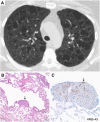
Figure 1.
Figure 1.
Lymphangio leiomyomatosis (LAM). Multiple smooth, round, thin-walled parenchymal cysts apparent on computed tomography imaging (A) correspond to histopathologic findings of parenchymal cystic spaces separated by normal intervening lung parenchyma (B) with focal aggregates of spindled and epithelioid LAM cells (B and C, arrows) with characteristic human melanoma black-45 (HMB-45) staining by immunohistochemistry (C, arrow, brown stain). Original magnifications: 10× (B); 40× (C).
Figure 2.
Figure 2.
Pulmonary Langerhans cell histiocytosis. Multiple nodules and cysts seen on computed tomography (CT) imaging (A) with histology showing cellular nodules (B), some with central cavities (B, *) containing diagnostic Langerhans cell aggregates highlighted by positive immunohistochemical staining for CD1a (C, brown stain) typical of the early cellular stage of the disease. Coronal CT image from another patient showing multiple bizarre-shaped cysts in an upper-zone–predominant distribution, with sparing of the costophrenic angles representative of later-stage disease (D). Histologic features typical of later disease stages include cystic spaces (E) associated with paucicellular stellate fibrosis (E, arrow). Accumulations of smoking-related pigmented macrophages (F, arrowhead) are frequently seen in the surrounding parenchyma. Original magnifications: 2× (E); 4× (B); 40× (C); 100× (F).
Figure 3.
Figure 3.
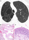
Cystic pleuropulmonary blastoma. Irregular, unilateral cysts apparent on computed tomography imaging (A) are characterized histologically as cysts (B) lined by a benign epithelium (B and C, arrows) with an underlying cambium layer of condensed immature mesenchymal cells (B and C, *). Original magnifications: 4× (B); 40× (C).
Figure 4.
Figure 4.
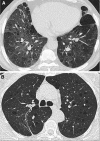
Diffuse cystic lung disease associated with smoke exposure. (A) Chest computed tomography (CT) showing thin-walled cysts interspersed with areas of ground-glass attenuation and paraseptal emphysema in a patient with desquamative interstitial pneumonia. (B) Chest CT showing round, thin-walled cysts with intervening normal lung parenchyma mimicking lymphangioleiomyomatosis in a patient with small airway damage secondary to cigarette smoke exposure.
Figure 5.
Figure 5.
Diffuse cystic lung disease associated with infectious etiologies. (A) Chest computed tomography (CT) showing multiple thick-walled cysts (pneumatoceles) and ground-glass opacities in a patient with Pneumocystis jiroveci. (B) Cytologic preparations of the bronchoalveolar lavage specimen showing frothy lavage fluid (*). (C) Fungal microorganisms are highlighted in the bronchoalveolar lavage specimen by Gomori methenamine silver (GMS) stain (arrow). (D) Chest CT showing thick-walled cysts along with nodules and ground-glass attenuation in a patient with recurrent respiratory papillomatosis. CT image of recurrent respiratory papillomatosis courtesy of Dr. Jonathan Chung (National Jewish Hospital, Denver, CO). Original magnifications: 100× (B and C).
Figure 6.
Figure 6.
Diffuse cystic lung disease associated with interstitial lung diseases. (A) Chest computed tomography (CT) showing multiple cysts of varying sizes in the left lower lobe associated with traction bronchiectasis, ground-glass attenuation, and areas of decreased lobar attenuation in a patient with chronic hypersensitivity pneumonitis. (B) CT showing cystic changes in a peripheral, subpleural distribution associated with reticulations and honeycombing in a patient with idiopathic pulmonary fibrosis.

References

    1. Hansell DM, Bankier AA, MacMahon H, McLoud TC, Müller NL, Remy J. Fleischner Society: glossary of terms for thoracic imaging. Radiology. 2008;246:697–722. - PubMed
    1. Henske EP, McCormack FX. Lymphangioleiomyomatosis - a wolf in sheep’s clothing. J Clin Invest. 2012;122:3807–3816. - PMC - PubMed
    1. McCormack FX, Travis WD, Colby TV, Henske EP, Moss J. Lymphangioleiomyomatosis: calling it what it is: a low-grade, destructive, metastasizing neoplasm. Am J Respir Crit Care Med. 2012;186:1210–1212. - PMC - PubMed
    1. Crino PB, Nathanson KL, Henske EP. The tuberous sclerosis complex. N Engl J Med. 2006;355:1345–1356. - PubMed
    1. Carsillo T, Astrinidis A, Henske EP. Mutations in the tuberous sclerosis complex gene TSC2 are a cause of sporadic pulmonary lymphangioleiomyomatosis. Proc Natl Acad Sci USA. 2000;97:6085–6090. - PMC - PubMed

MeSH terms

Supplementary concepts