

Amyloid burden in the hippocampus and default mode network: relationships with gray matter volume and cognitive performance in mild stage Alzheimer disease
- PMID: 25906109
- PMCID: PMC4602683
- DOI: 10.1097/MD.0000000000000763
Amyloid burden in the hippocampus and default mode network: relationships with gray matter volume and cognitive performance in mild stage Alzheimer disease
Abstract
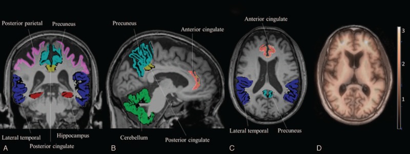
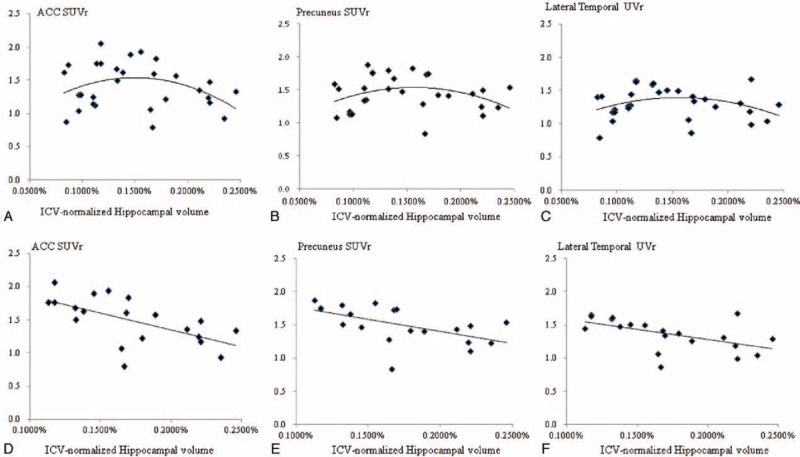
Amyloid load, as measured by florbetapir positron emission tomography (PET) standardized uptake value ratio (SUVr), has high specificity in the diagnosis of Alzheimer disease (AD). As the posterior cingulate cortex (PCC) represents densely amyloid-affected regions early in AD, we hypothesized that amyloid load within the key hubs of the default mode networks (DMN) may result in local or distant interconnected gray matter (GM) volume atrophy, thereby affecting cognitive performance. Thirty AD patients with a clinical dementia rating sum of box score ≤2 were enrolled and underwent cognitive evaluation, 3-dimensional T1-weighted imaging and florbetapir PET. Volumes of interest (VOIs) included the hippocampus, lateral temporal region, and key hubs of the DMN [anterior cingulate cortex (ACC), PCC, posterior parietal, and precuneus]. The SUVr was calculated by florbetapir standard uptake value (SUV) within the T1-weighted image segmented GM VOIs divided by the cerebellar GM SUV. Our results suggested inverse correlations between ACC (ρ = -0.444, P = 0.016) and PCC SUVr (ρ = -0.443, P = 0.016) with PCC GM volume. In stepwise regression, the orientation scores were associated with PCC SUVr (β = 2.584, P = 0.02) and posterior parietal volume (β = -0.446, P = 0.04), whereas the word recall score was related to hippocampal volume (β = -0.391, P = 0.04). After removing the patients with a hippocampal VOI below the lowest tertile and adjusting for age, an inverse correlation was found between hippocampal volume and SUVr in the ACC (partial σ = -0.639, P = 0.002), precuneus (partial σ = -0.692, P = 0.002), and lateral temporal SUVr (partial σ = -0.604, P = 0.005). Our results suggest that amyloid burden within the key DMN regions may contribute to local and distant GM atrophy, and that this may explain the cognitive scores.
Conflict of interest statement
The authors have no conflicts of interest to disclose.
Figures
References
-
- Dubois B, Feldman HH, Jacova C, et al. Advancing research diagnostic criteria for Alzheimer's disease: the IWG-2 criteria. Lancet Neurol 2014; 13:614–629. - PubMed
-
- Chetelat G, Villemagne VL, Bourgeat P, et al. Relationship between atrophy and beta-amyloid deposition in Alzheimer disease. Ann Neurol 2010; 67:317–324. - PubMed
Publication types
MeSH terms
Substances
LinkOut - more resources
Full Text Sources
Medical
Research Materials
