

High prevalence of hepatitis e virus in Swedish moose--a phylogenetic characterization and comparison of the virus from different regions
- PMID: 25906163
- PMCID: PMC4408071
- DOI: 10.1371/journal.pone.0122102
High prevalence of hepatitis e virus in Swedish moose--a phylogenetic characterization and comparison of the virus from different regions
Abstract
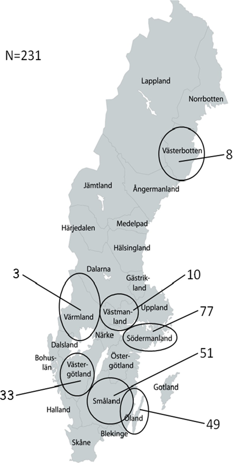
Background: Hepatitis E virus (HEV) infects a range of species, including humans, pigs, wild boars and deer. Zoonotic transmission may contribute to the high HEV seroprevalence in the human population of many countries. A novel divergent HEV from moose (Alces alces) in Sweden was recently identified by partial genome sequencing. Since only one strain was found, its classification within the HEV family, prevalence in moose and zoonotic potential was unclear. We therefore investigated samples from 231 moose in seven Swedish counties for HEV, and sequenced a near complete moose HEV genome. Phylogenetic analysis to classify this virus within the family Hepeviridae and to explore potential host specific determinants was performed.
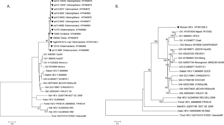
Methods and findings: The HEV prevalence of moose was determined by PCR (marker for active infection) and serological assays (marker of past infection) of sera and 51 fecal samples from 231 Swedish moose. Markers of active and past infection were found in 67 (29%) animals, while 34 (15%) were positive for HEV RNA, 43 (19%) were seropositive for anti-HEV antibodies, and 10 (4%) had both markers. The number of young individuals positive for HEV RNA was larger than for older individuals, and the number of anti-HEV antibody positive individuals increased with age. The high throughput sequenced moose HEV genome was 35-60% identical to existing HEVs. Partial ORF1 sequences from 13 moose strains showed high similarity among them, forming a distinct monophyletic clade with a common ancestor to HEV genotype 1-6 group, which includes members known for zoonotic transmission.
Conclusions: This study demonstrates a high frequency of HEV in moose in Sweden, with markers of current and past infection demonstrated in 30% of the animals. Moose is thus an important animal reservoir of HEV. The phylogenetic relationship demonstrated that the moose HEV belonged to the genotype 1-6 group, which includes strains that also infect humans, and therefore may signify a potential for zoonotic transmission of this HEV.
Conflict of interest statement
Figures
References
Publication types
MeSH terms
Substances
LinkOut - more resources
Full Text Sources
Other Literature Sources
Miscellaneous
