Review
. 2015 Jul 1;192(1):17-29.
doi: 10.1164/rccm.201411-2096CI.

Diffuse Cystic Lung Disease. Part II

Affiliations
Review

Diffuse Cystic Lung Disease. Part II

Nishant Gupta et al. Am J Respir Crit Care Med. .

Abstract

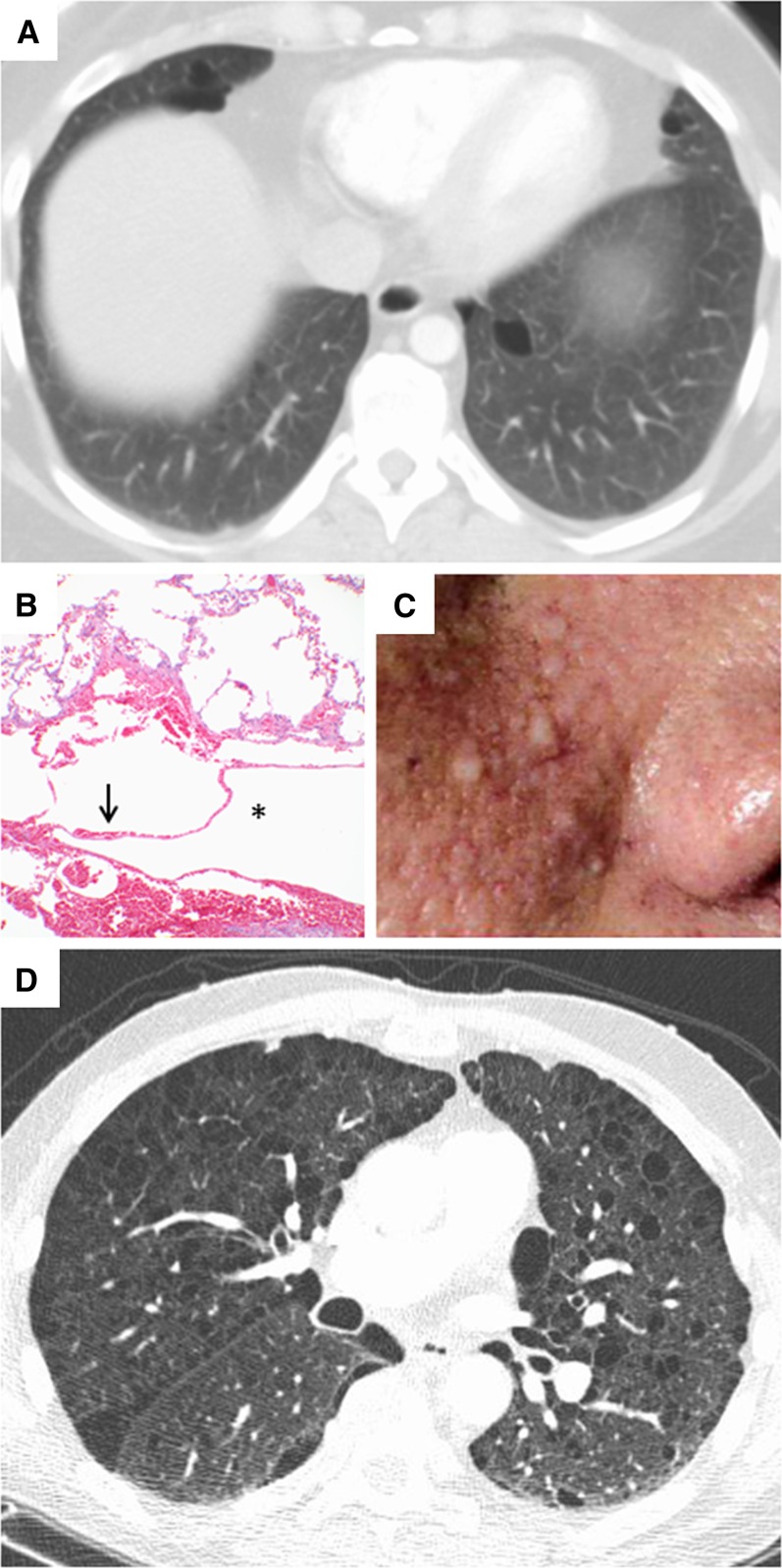
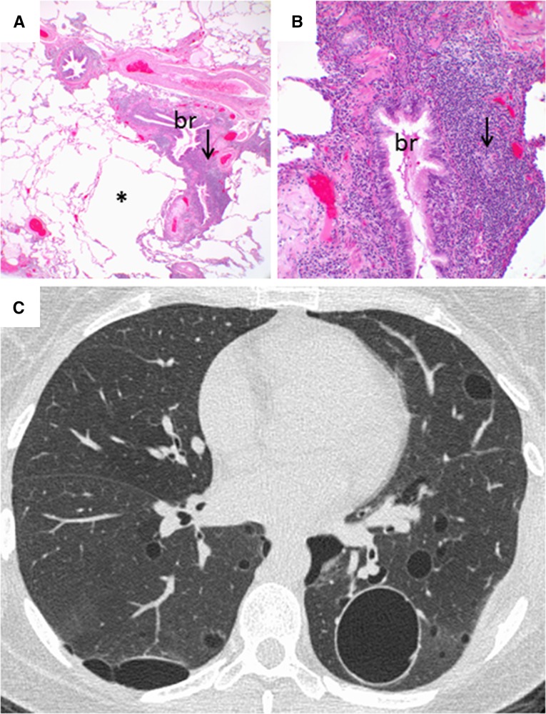
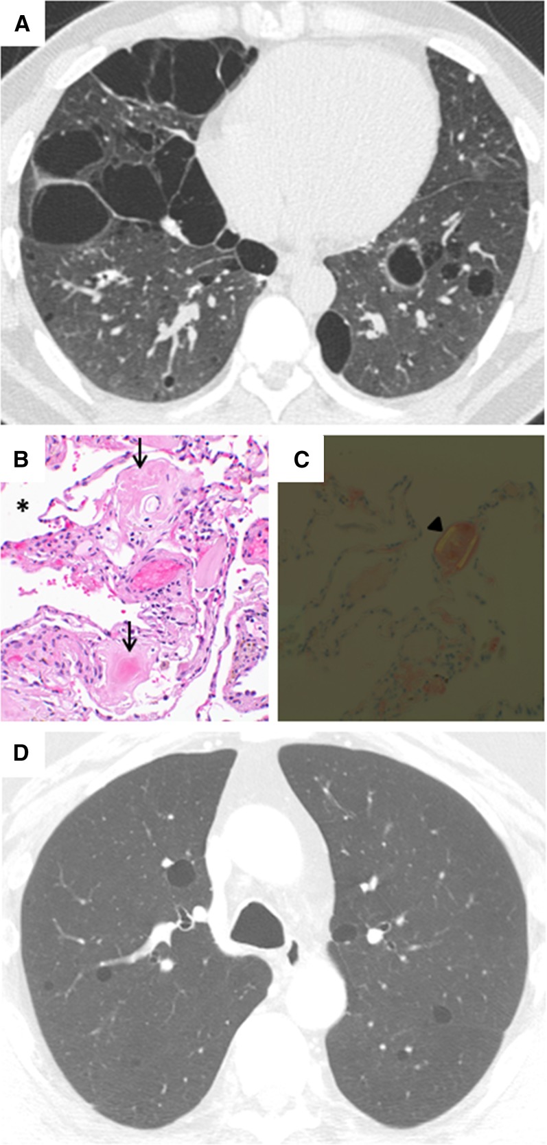
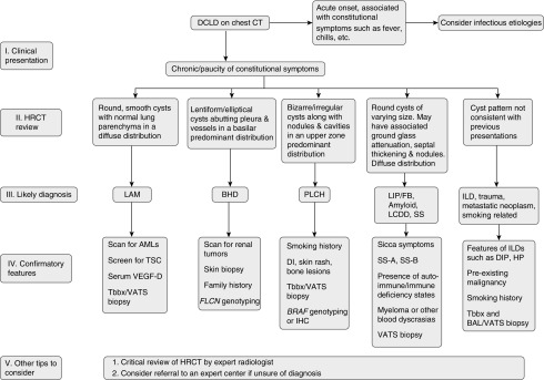
The diffuse cystic lung diseases have a broad differential diagnosis. A wide variety of pathophysiological processes spanning the spectrum from airway obstruction to lung remodeling can lead to multifocal cyst development in the lung. Although lymphangioleiomyomatosis and pulmonary Langerhans cell histiocytosis are perhaps more frequently seen in the clinic, disorders such as Birt-Hogg-Dubé syndrome, lymphocytic interstitial pneumonia, follicular bronchiolitis, and light-chain deposition disease are increasingly being recognized. Obtaining an accurate diagnosis can be challenging, and management approaches are highly disease dependent. Unique imaging features, genetic tests, serum studies, and clinical features provide invaluable clues that help clinicians distinguish among the various etiologies, but biopsy is often required for definitive diagnosis. In part II of this review, we present an overview of the diffuse cystic lung diseases caused by lymphoproliferative disorders, genetic mutations, or aberrant lung development and provide an approach to aid in their diagnosis and management.

Keywords: Birt-Hogg-Dubé syndrome; Sjögren syndrome; follicular bronchiolitis; high-resolution computed tomography; lymphoid interstitial pneumonia.


Figures

Figure 1.
Figure 1.
Computed tomography (CT) and histopathologic images of diffuse cystic lung disease associated with genetic mutations. (A) Chest CT showing lentiform cysts in a basilar distribution in a patient with Birt-Hogg-Dubé (BHD) syndrome. (B) Histologic sections of a lung biopsy from a patient with BHD syndrome showing intraparenchymal cysts (*) surrounded by normal parenchyma with an intracystic septum (arrow) and lacking abnormal cellular proliferations or a significant fibroinflammatory component. Original magnification, ×10. (C) Facial photograph of a patient with BHD syndrome showing multiple dome-shaped, whitish papules consistent with fibrofolliculomas in the nasolabial fold area. (D) Chest CT showing multiple thin-walled cysts and paraseptal emphysema in a patient with neurofibromatosis (courtesy of Dr. Cristopher Meyer, University of Wisconsin, Madison, WI).
Figure 2.
Figure 2.
Computed tomography and histopathologic images of follicular bronchiolitis. (A) Histologic sections showing parenchymal cysts (*) associated with follicular lymphoid hyperplasia (arrow) centered around bronchioles (br). Original magnification, ×2. (B) Higher-power view of histologic image in A showing follicular hyperplasia (arrow) surrounding the bronchiole (br). Original magnification, ×20. (C) Chest computed tomography showing multiple cysts of varying sizes associated with minimal ground-glass opacification.
Figure 3.
Figure 3.
Computed tomography (CT) and histopathologic images of diffuse cystic lung disease associated with lymphoproliferative disorders with protein deposition. (A) Chest CT showing diffuse, irregular cysts in a patient with amyloidosis. (B) Histologic sections of a lung biopsy from a patient with amyloidosis showing intraparenchymal cysts (*) and accumulation of amyloid (arrows) in the surrounding parenchyma and vessels. Original magnification, ×40. (C) The amyloid deposition in this biopsy shows the characteristic apple-green birefringence under polarized light (arrowhead). Original magnification, ×40. (D) Chest CT showing multiple round, thin-walled cysts in a patient with light-chain deposition disease (courtesy of Dr. Jonathan Chung, National Jewish Health, Denver, CO).
Figure 4.
Figure 4.
Algorithm to guide approach to the diagnosis of diffuse cystic lung diseases. AML = angiomyolipoma; BAL = bronchioalveolar lavage; BHD = Birt-Hogg-Dubé syndrome; BRAF = v-Raf murine sarcoma viral oncogene homolog B; CT = computed tomography; DCLD = diffuse cystic lung disease; DI = diabetes insipidus; DIP = desquamative interstitial pneumonia; FB = follicular bronchiolitis; FLCN = folliculin; HRCT = high-resolution computed tomography; HP = hypersensitivity pneumonitis; IHC = immunohistochemistry; ILD = interstitial lung disease; LAM = lymphangioleiomyomatosis; LCDD = light-chain deposition disease; LIP = lymphoid interstitial pneumonia; PLCH = pulmonary Langerhans cell histiocytosis; SS = Sjögren syndrome; Tbbx = transbronchial biopsy; TSC = tuberous sclerosis complex; VATS = video-assisted thoracoscopic surgery; VEGF-D = vascular endothelial growth factor-D.

References

    1. Kunogi M, Kurihara M, Ikegami TS, Kobayashi T, Shindo N, Kumasaka T, Gunji Y, Kikkawa M, Iwakami S, Hino O, et al. Clinical and genetic spectrum of Birt-Hogg-Dube syndrome patients in whom pneumothorax and/or multiple lung cysts are the presenting feature. J Med Genet. 2010;47:281–287. - PMC - PubMed
    1. Gunji Y, Akiyoshi T, Sato T, Kurihara M, Tominaga S, Takahashi K, Seyama K. Mutations of the Birt Hogg Dube gene in patients with multiple lung cysts and recurrent pneumothorax. J Med Genet. 2007;44:588–593. - PMC - PubMed
    1. Tomassetti S, Carloni A, Chilosi M, Maffè A, Ungari S, Sverzellati N, Gurioli C, Casoni G, Romagnoli M, Gurioli C, et al. Pulmonary features of Birt-Hogg-Dubé syndrome: cystic lesions and pulmonary histiocytoma. Respir Med. 2011;105:768–774. - PubMed
    1. Toro JR, Pautler SE, Stewart L, Glenn GM, Weinreich M, Toure O, Wei MH, Schmidt LS, Davis L, Zbar B, et al. Lung cysts, spontaneous pneumothorax, and genetic associations in 89 families with Birt-Hogg-Dubé syndrome. Am J Respir Crit Care Med. 2007;175:1044–1053. - PMC - PubMed
    1. Onuki T, Goto Y, Kuramochi M, Inagaki M, Bhunchet E, Suzuki K, Tanaka R, Furuya M. Radiologically indeterminate pulmonary cysts in Birt-Hogg-Dubé syndrome. Ann Thorac Surg. 2014;97:682–685. - PubMed

MeSH terms

Supplementary concepts