. 2015 Jul 1;192(1):30-9.
doi: 10.1164/rccm.201412-2293OC.

Morphologic Characterization of Nerves in Whole-Mount Airway Biopsies

Affiliations

Morphologic Characterization of Nerves in Whole-Mount Airway Biopsies

Peter W West et al. Am J Respir Crit Care Med. .

Abstract

Rationale: Neuroplasticity of bronchopulmonary afferent neurons that respond to mechanical and chemical stimuli may sensitize the cough reflex. Afferent drive in cough is carried by the vagus nerve, and vagal afferent nerve terminals have been well defined in animals. Yet, both unmyelinated C fibers and particularly the morphologically distinct, myelinated, nodose-derived mechanoreceptors described in animals are poorly characterized in humans. To date there are no distinctive molecular markers or detailed morphologies available for human bronchopulmonary afferent nerves.

Objectives: Morphologic and neuromolecular characterization of the afferent nerves that are potentially involved in cough in humans.

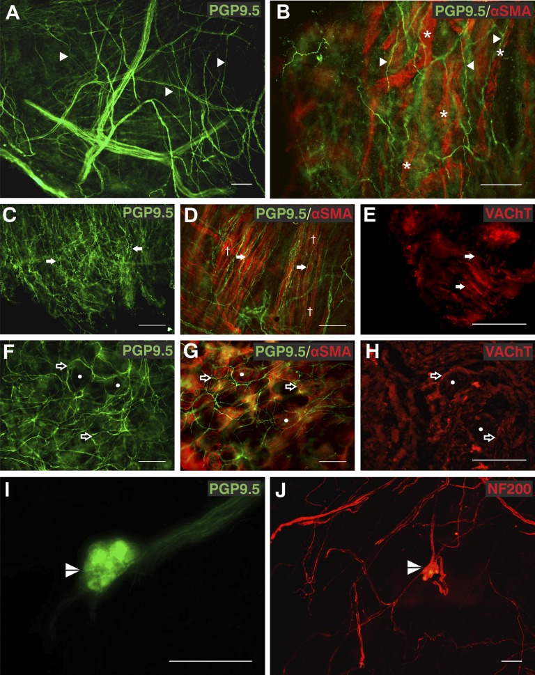
Methods: A whole-mount immunofluorescence approach, rarely used in human lung tissue, was used with antibodies specific to protein gene product 9.5 (PGP9.5) and, for the first time in human lung tissue, 200-kD neurofilament subunit.

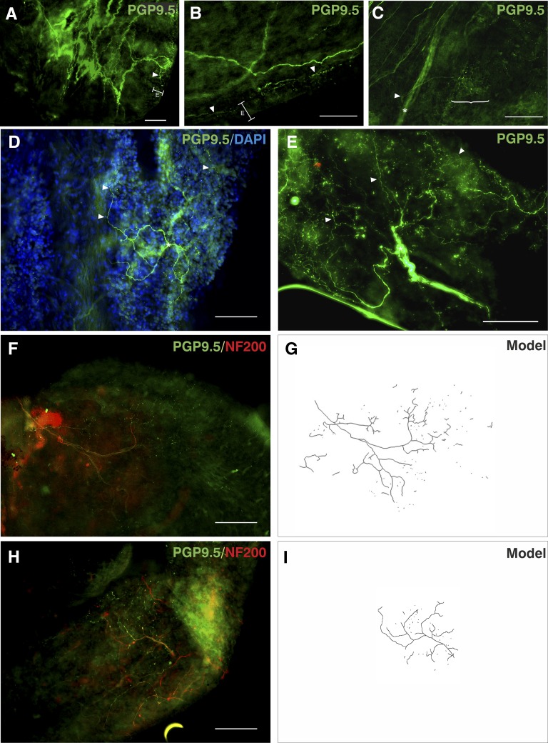
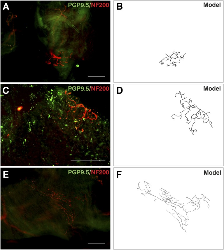
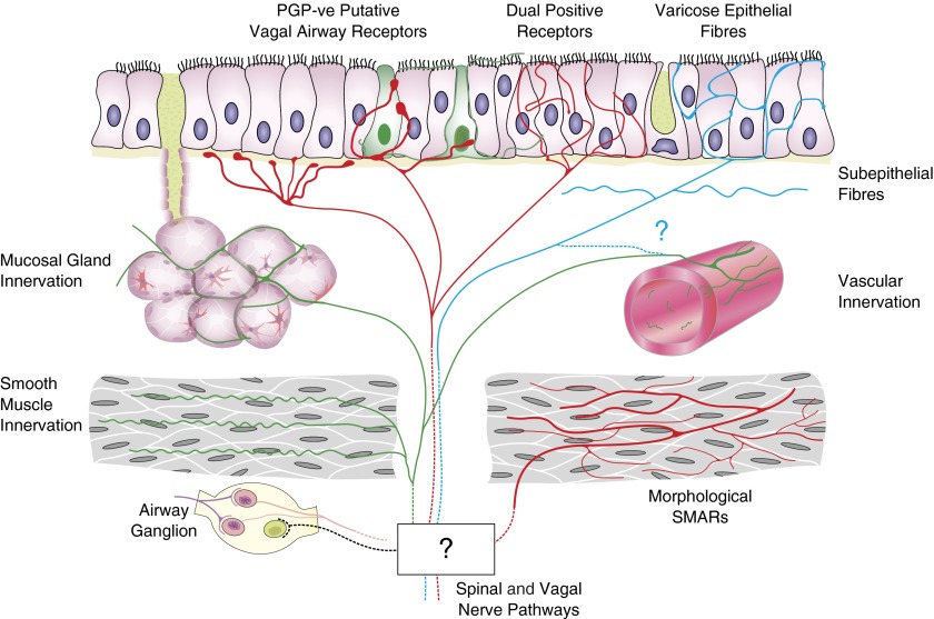
Measurements and main results: We have developed a robust technique to visualize fibers consistent with autonomic and C fibers and pulmonary neuroendocrine cells. A group of morphologically distinct, 200-kD neurofilament-immunopositive myelinated afferent fibers, a subpopulation of which did not express PGP9.5, was also identified.

Conclusions: PGP9.5-immunonegative nerves are strikingly similar to myelinated airway afferents, the cough receptor, and smooth muscle-associated airway receptors described in rodents. These have never been described in humans. Full description of human airway nerves is critical to the translation of animal studies to the clinical setting.

Keywords: afferent neurons; lung; mechanoreceptors; peripheral nervous system.


Figures

Figure 1.
Figure 1.
Autonomic fibers in the human airway mucosa. (A and B) Protein gene product 9.5 (PGP9.5) immunopositive (green, Alexa Fluor 488) subepithelial fibers (arrowheads) were associated with α-smooth muscle actin (SMA)-immunoreactive (red, Alexa Fluor 568) blood vessels (asterisks). (C–E) PGP9.5-immunopositive (green, Alexa Fluor 488) convoluted fibers (arrows) were observed in parallel to bronchial smooth muscle (daggers) labeled with α-SMA (red, Alexa Fluor 568) and a proportion were immunopositive for vesicular acetylcholine transporter (VAChT) (red, Alexa Fluor 568). (F–H) The distinctive circular pattern of PGP9.5 and VAChT immunoreactive fibers (open arrows) around acinar cells of bronchial mucosal glands observed with α-SMA (circles). (I and J) Airway intrinsic autonomic ganglia (split arrowheads) immunoreactive for both PGP9.5 (green, Alexa Fluor 488) and 200-kD neurofilament (NF200; red, Alexa Fluor 568) were observed. Biopsies were from right middle lobe entrance (A, B, E, F, and H), right lower lobe (C, D, and G), right upper lobe entrance (I), and right main bronchus (J). Results are independent experiments of staining in nine biopsies from seven patients. Scale bars = 100 μm. Unprocessed and single-channel images are shown in Figures E1 and E2 with additional images in Figure E3. A video of additional images of separate focal planes from B is shown in Video E1.
Figure 2.
Figure 2.
Protein gene product 9.5 (PGP9.5) immunoreactivity in human airway epithelium. (A and B) PGP9.5-immunopositive (green, Alexa Fluor 488) varicose epithelial fibers (arrowheads) appeared in patches at the luminal edge of the epithelium. (C and D) A population of epithelial cells were immunoreactive for PGP9.5, with typical flask-shaped pulmonary neuroendocrine cell morphology (asterisks) and cellular projections (open arrows). Segments of epithelial fibers were also observed (arrowheads). (E and F) Epithelial fibers were mostly 200-kD neurofilament (NF200) immunonegative (open arrow), but some were observed only with antineurofilament (red, Alexa Fluor 568, solid arrow) or were dual positive (arrowheads). Biopsies were from right main bronchus (A, D, and E) and right middle lobe entrance (B, C, and F). Results shown are independent experiments of six biopsies from separate patients. Scale bars = 100 μm. Unprocessed, single-channel images, additional images, and videos are shown in Figures E4–E6 and Videos E2 and E3.
Figure 3.
Figure 3.
Nerve fiber diameter analysis. The median fiber diameter (in micrometers) of five identified protein gene product 9.5–stained fiber subtypes is shown. Measurements (n = 200 fibers) were obtained from 31 biopsies of 18 patients and grouped morphometrically into epithelial (n = 35), subepithelial (n = 42), mucosal gland (n = 32), convoluted (n = 44), and nerve trunk (n = 40). Individual fiber diameters are represented as separate data points, with midline and error bars to indicate median diameters with interquartile range. ***Significant differences (P < 0.001, Kruskal-Wallis test with Dunn’s post-test correction).
Figure 4.
Figure 4.
Protein gene product 9.5 (PGP9.5)-immunoreactive putative airway afferent fibers. Immunostaining for PGP9.5 (green, Alexa Fluor 488) 200-kD neurofilament (NF200; red, Alexa Fluor 568) using 4′,6-diamidino-2-phenylindole (DAPI; blue) as a nuclear counterstain (where shown). (A and B) Nerves that gave rise to varicosities in the epithelial plexus (E) were associated with nerve branches located superficially near the epithelial basement membrane (arrowheads). (C) Some similar-sized axons (arrowhead), which gave rise to epithelial fibers (bracket), arose directly from deeper nerve trunks (asterisk). (D and E) Varicose epithelial fibers (arrowheads) were arranged into extensively branched arboreal structures from a central point (asterisks). (F–I) The main branches of these fibers were immunopositive for NF200, whereas varicose nerve endings were only immunopositive for PGP9.5. (G and I) Computerized tracings show that these fibers form part of a larger arboreal structure, which appeared partly fragmented. Data are from biopsies from right middle lobe entrance (A–C and H); right lower lobe (D and E); and right main bronchus (F) from six separate patients. E is a maximum-intensity projection of five epifluorescent images. Scale bars = 100 μm. Unprocessed, single-channel, and additional images are shown in Figures E7 and E8 and Video E4.
Figure 5.
Figure 5.
Protein gene product 9.5 (PGP9.5)-immunonegative afferent fibers. Tissue was observed using PGP9.5 (green, Alexa Fluor 488) and 200-kD neurofilament (NF200; red, Alexa Fluor 568) immunofluroescence. (A, C, and E) Neuronal structures were observed that were PGP9.5 negative but NF200 positive. There were fewer other neuronal structures in these regions, but successful PGP9.5 immunostaining identified neurons and pulmonary neuroendocrine cells (A and C). Pulmonary neuroendocrine cells were observed in close association with terminals of a thick NF200-stained nerve. (B, D, and F) Computerized grayscale tracings show the outline structure of fibers. Data are from three biopsies from the right middle lobe entrance (A and C) and right main bronchus (E) of three separate subjects. Scale bars = 200 μm. Unprocessed, single-channel, and additional images are shown in Figures E9–E11 and Video E5.
Figure 6.
Figure 6.
Schematic representation of the putative innervation of human airway mucosa based on morphologic observations from this study. Areas that are unknown or unproved are notated by a question mark. Airway ganglia and protein gene product 9.5 (PGP9.5)-immunopositive innervation of mucosal glands, smooth muscle, and mucosal blood vessels was observed. Putative afferent epithelial fibers with multiple morphologies may represent separate types. PGP9.5-immunonegative (PGP-ve) putative vagal afferents and morphologic equivalents of rodent vagal afferent receptors were identified with antibodies to 200-kD neurofilament. Varicose epithelial fibers have morphology and diameters consistent with C fibers. Neurofilament-immunopositive fine epithelial fibers may be the terminals of myelinated afferents. SMAR = smooth muscle–associated airway receptor.

Comment in

References

    1. Canning BJ, Woo A, Mazzone SB. Neuronal modulation of airway and vascular tone and their influence on nonspecific airways responsiveness in asthma. J Allergy (Cairo) 2012;2012:108149. - PMC - PubMed
    1. Voll-Aanerud M, Eagan TML, Plana E, Omenaas ER, Bakke PS, Svanes C, Siroux V, Pin I, Antó JM, Leynaert B. Respiratory symptoms in adults are related to impaired quality of life, regardless of asthma and COPD: results from the European community respiratory health survey. Health Qual Life Outcomes. 2010;8:107. - PMC - PubMed
    1. Irwin RS, Baumann MH, Bolser DC, Boulet LP, Braman SS, Brightling CE, Brown KK, Canning BJ, Chang AB, Dicpinigaitis PV, et al. American College of Chest Physicians (ACCP) Diagnosis and management of cough executive summary: ACCP evidence-based clinical practice guidelines. Chest. 2006;129(1, Suppl):1S–23S. - PMC - PubMed
    1. Prudon B, Birring SS, Vara DD, Hall AP, Thompson JP, Pavord ID. Cough and glottic-stop reflex sensitivity in health and disease. Chest. 2005;127:550–557. - PubMed
    1. Groneberg DA, Niimi A, Dinh QT, Cosio B, Hew M, Fischer A, Chung KF. Increased expression of transient receptor potential vanilloid-1 in airway nerves of chronic cough. Am J Respir Crit Care Med. 2004;170:1276–1280. - PubMed

Publication types

MeSH terms

LinkOut - more resources