

Multivariate morphological brain signatures predict patients with chronic abdominal pain from healthy control subjects
- PMID: 25906347
- PMCID: PMC4504800
- DOI: 10.1097/j.pain.0000000000000196
Multivariate morphological brain signatures predict patients with chronic abdominal pain from healthy control subjects
Abstract
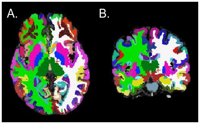
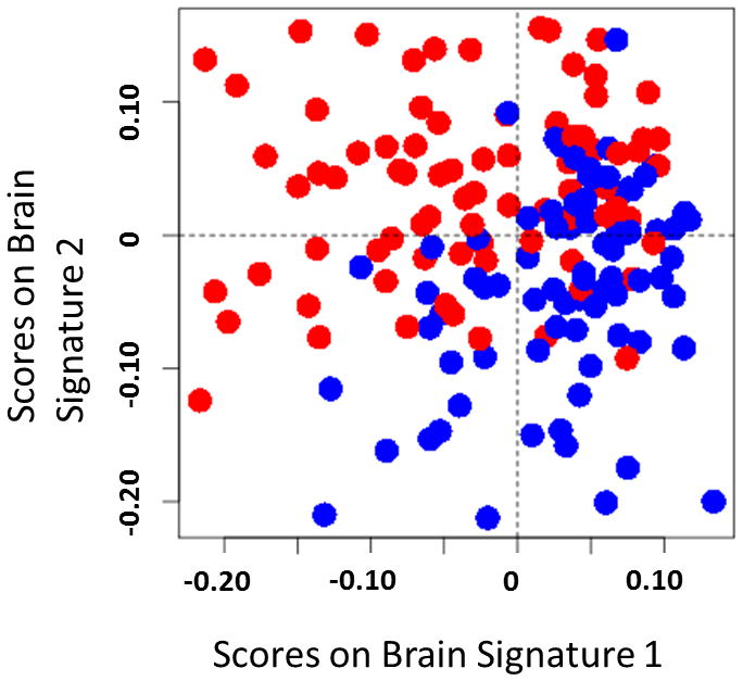
Irritable bowel syndrome (IBS) is the most common chronic visceral pain disorder. The pathophysiology of IBS is incompletely understood; however, evidence strongly suggests dysregulation of the brain-gut axis. The aim of this study was to apply multivariate pattern analysis to identify an IBS-related morphometric brain signature that could serve as a central biological marker and provide new mechanistic insights into the pathophysiology of IBS. Parcellation of 165 cortical and subcortical regions was performed using FreeSurfer and the Destrieux and Harvard-Oxford atlases. Volume, mean curvature, surface area, and cortical thickness were calculated for each region. Sparse partial least squares discriminant analysis was applied to develop a diagnostic model using a training set of 160 females (80 healthy controls and 80 patients with IBS). Predictive accuracy was assessed in an age-matched holdout test set of 52 females (26 healthy controls and 26 patients with IBS). A 2-component classification algorithm comprising the morphometry of (1) primary somatosensory and motor regions and (2) multimodal network regions explained 36% of the variance. Overall predictive accuracy of the classification algorithm was 70%. Small effect size associations were observed between the somatosensory and motor signature and nongastrointestinal somatic symptoms. The findings demonstrate that the predictive accuracy of a classification algorithm based solely on regional brain morphometry is not sufficient, but they do provide support for the utility of multivariate pattern analysis for identifying meaningful neurobiological markers in IBS.
Conflict of interest statement
The authors report no conflict of interest.
Figures
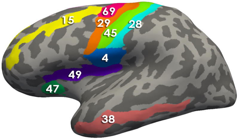
Brain Signature 1 regions-Lateral View: 4, Subcentral Gyrus (central operculum) and Sulci; 15, Middle Frontal Gyrus (15); 28, Postcentral Gyrus; 29, Precentral Gyrus; 38, Middle Temporal Gyrus; 45, Central Sulcus (Rolando’s Fissure); 47, Anterior Segment of the Circular Sulcus of the Insula; 49, Superior Segment of the Circular Sulcus of the Insula; 69, Superior Part of the Precentral SulcusBrain Signature 1 regions-Medial View : 3, Paracentral Lobule and Sulcus; 8, Middle-Posterior Part of the Cingulate Gyrus and Sulcus (pMCC); 16, Superior Frontal Gyrus 66, Pericallosal Sulcus (Sulcus of Corpus Callosum); 70, Suborbital Sulcus (Sulcus Rostrales, Supraorbital Sulcus)
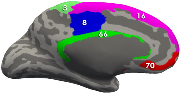
Brain Signature 1 regions-Lateral View: 4, Subcentral Gyrus (central operculum) and Sulci; 15, Middle Frontal Gyrus (15); 28, Postcentral Gyrus; 29, Precentral Gyrus; 38, Middle Temporal Gyrus; 45, Central Sulcus (Rolando’s Fissure); 47, Anterior Segment of the Circular Sulcus of the Insula; 49, Superior Segment of the Circular Sulcus of the Insula; 69, Superior Part of the Precentral SulcusBrain Signature 1 regions-Medial View : 3, Paracentral Lobule and Sulcus; 8, Middle-Posterior Part of the Cingulate Gyrus and Sulcus (pMCC); 16, Superior Frontal Gyrus 66, Pericallosal Sulcus (Sulcus of Corpus Callosum); 70, Suborbital Sulcus (Sulcus Rostrales, Supraorbital Sulcus)
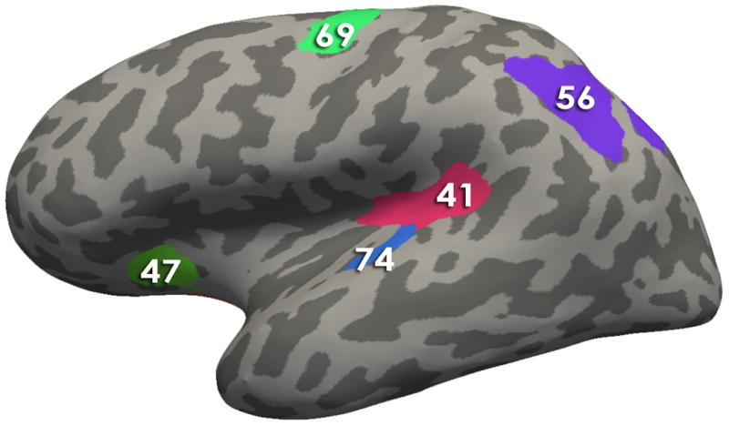
Brain Signature 2 regions-Lateral View: 41, Posterior Ramus of the Lateral Sulcus; 47, Anterior Segment of the Circular Sulcus of the Insula; 69, Superior Part of the Precentral Sulcus; 56, Intraparietal Sulcus and Transverse Parietal Sulci ; 74, Transverse Temporal Sulcus
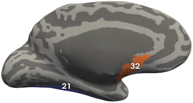
Brain Signature 2 regions- Medial View: 21 Lateral Occipito-Temporal Gyrus (Fusiform Gyrus); 32, Subcallosal Area (Subcallosal Gyrus);
Brain Signature 2 regions-Lateral View: 41, Posterior Ramus of the Lateral Sulcus; 47, Anterior Segment of the Circular Sulcus of the Insula; 69, Superior Part of the Precentral Sulcus; 56, Intraparietal Sulcus and Transverse Parietal Sulci ; 74, Transverse Temporal Sulcus
Brain Signature 2 regions- Medial View: 21 Lateral Occipito-Temporal Gyrus (Fusiform Gyrus); 32, Subcallosal Area (Subcallosal Gyrus);
Comment in
-
Neuroimaging-based biomarker discovery and validation.Pain. 2015 Aug;156(8):1379-1381. doi: 10.1097/j.pain.0000000000000223. Pain. 2015. PMID: 25970320 Free PMC article. No abstract available.
References
-
- Pain and Interoception Imaging Network. 2015
-
- Andresen V, Bach DR, Poellinger A, Tsrouya C, Stroh A, Foerschler A, Georgiewa P, Zimmer C, Monnikes H. Brain activation responses to subliminal or supraliminal rectal stimuli and to auditory stimuli in irritable bowel syndrome. Neurogastroenterology and motility : the official journal of the European Gastrointestinal Motility Society. 2005;17(6):827–837. - PubMed
-
- Andresen V, Poellinger A, Tsrouya C, Bach D, Stroh A, Foerschler A, Georgiewa P, Schmidtmann M, van der Voort IR, Kobelt P, Zimmer C, Wiedenmann B, Klapp BF, Monnikes H. Cerebral processing of auditory stimuli in patients with irritable bowel syndrome. World journal of gastroenterology : WJG. 2006;12(11):1723–1729. - PMC - PubMed
-
- Berman SM, Naliboff BD, Chang L, Fitzgerald L, Antolin T, Camplone A, Mayer EA. Enhanced preattentive central nervous system reactivity in irritable bowel syndrome. The American journal of gastroenterology. 2002;97(11):2791–2797. - PubMed
Publication types
MeSH terms
Substances
Grants and funding
LinkOut - more resources
Full Text Sources
Other Literature Sources
Medical
