Review
. 2015 Apr 25:13:163.
doi: 10.1186/s12957-015-0573-y.

Primary tumors of the patella

Affiliations
Review

Primary tumors of the patella

Mingzhi Song et al. World J Surg Oncol. .

Abstract

The patella is an uncommon location for cancerous occurrence and development. The majority of tumors of the patella are benign, with a significant incidence of giant cell tumors and chondroblastoma. With the development of modern diagnostic technologies, there appear however many other histological types which raise challenges of diagnosis and treatment. In this article, we review the reported histological types of primary patellar tumors. Specifically, epidemiology, symptomatology, imageology, histopathology, and treatment options for these histological lesions will be discussed, respectively. As there is an increasing focus on the diagnosis and the treatment of these lesions, the availability of the integrated information about primary patellar tumors becomes more significant.


Figures

Figure 1
Figure 1
Patellar giant cell tumor. (A, B) An enlargement of the cystic lesion in the patella and a bone translucency with peripheral rim change can be seen at the lateral down part of the patella on radiographs. (C, D) CT indicates that there is a well-defined lytic lesion with the thin cortex occupying two-thirds of the patella.
Figure 2
Figure 2
Chondroblastomas of the patella. (A) On plain radiographs, the lesion appears as geographic changes with lobulated margins, thinning cortex, sclerotic rim, and reactive bone. (B) Calcifications inside the lesion are seen on CT images.
Figure 3
Figure 3
An intracortical osteoid osteoma of the patella. (A) On plain radiographs, a clear lesion with small fuzzy periosteal reaction is observed. (B) CT demonstrates the sclerosis surrounding the lesion.
Figure 4
Figure 4
Patellar hemangioma. (A) Radiographs show a lytic lesion with sharp margins, thinning cortex, and sclerotic rim. (B, C) CT results accord with the radiographic results.
Figure 5
Figure 5
Osteosarcoma of the patella. (A) Plain radiograph and (B, C) CT images reveal a lytic lesion in the patella with partial cortical disruption.
Figure 6
Figure 6
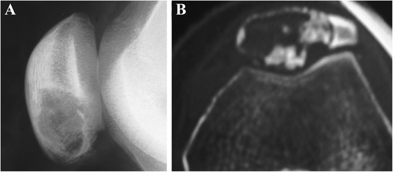
Patellar lymphoma with a pathologic fracture. (A) On radiographs, the patella consists of a sclerotic proximal portion and a lytic distal portion. (B) Permeative bone destruction, hazy margins, destroyed cortex, large soft tissue mass surrounding the patella, and joint involvement are shown on CT images.
Figure 7
Figure 7
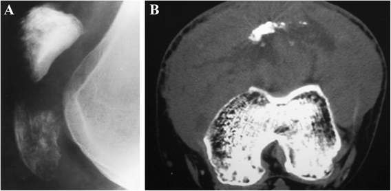
Patellar leiomyosarcoma. (A) Plain radiograph shows a mixed lytic and sclerotic lesion of patella. The margin of the lesion is ill defined and associated with cortical breach. (B) CT scan reveals multiple lytic lesions of the patella with a sclerotic rim and cortical disruption.
Figure 8
Figure 8
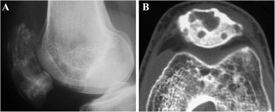
Hemangioendothelioma of the patella. Septation and thinning cortex in the osteolytic lesion are observed on (A) plain radiographs and (B) CT images.

References

    1. Mercuri M, Casadei R. Patellar tumors. Clin Orthop Relat. 2001;163:35–46. doi: 10.1097/00003086-200108000-00007. - DOI - PubMed
    1. Yoshida Y, Kojima T, Taniguchi M, Osaka S, Tokuhashi Y. Giant-cell tumor of the patella. Acta Med Okayama. 2012;66:73–6. - PubMed
    1. Ofluoglu O, Donthineni R. Iatrogenic seeding of a giant cell tumor of the patella to the proximal tibia. Clin Orthop Relat Res. 2007;465:260–4. - PubMed
    1. Malhotra R, Sharma L, Kumar V, Nataraj AR. Giant cell tumor of the patella and its management using a patella, patellar tendon, and tibial tubercle allograft. Knee Surg Sports Traumatol Arthrosc. 2010;18:167–9. doi: 10.1007/s00167-009-0908-8. - DOI - PubMed
    1. Chakraverty G, Chakraverty U. Late presentation of G.C.T. of the patella. Internet J Orthopedic. Surgery. 2009;12:1.