

Distal femoral condyle is more internally rotated to the patellar tendon at 90° of flexion in normal knees
- PMID: 25906977
- PMCID: PMC4410736
- DOI: 10.1186/s13018-015-0197-5
Distal femoral condyle is more internally rotated to the patellar tendon at 90° of flexion in normal knees
Abstract
Background: The configuration of the distal surface of the femur would be more important in terms of the patellofemoral (PF) joint contact because the patella generally contacts with the distal surface of the femur in knee flexion. Some total knee arthroplasty (TKA) designs configurate medially prominent asymmetric femoral condyles. This difference in the design of distal femoral condyle may affect the PF joint congruity in knee flexion. Furthermore, some surgeons advocate a concept aligning the symmetric components parallel to the native joint inclination, not perpendicular to the mechanical axis. This concept would also make a difference on the PF joint congruity at the distal femur in knee flexion. However, no fundamental study has been reported on the PF congruity at the distal femur to discuss the theoretical priority of these concepts. The current study investigated the angular relationship between the tibial attachment of the patellar tendon and the distal surface of the femur at 90° of flexion in normal knees.
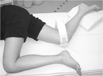
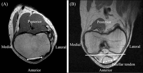
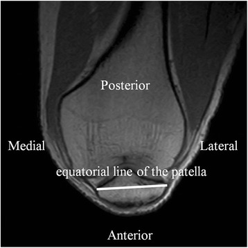
Methods: The open magnetic resonance images of 45 normal knees at 90° of flexion were used to measure the angles between the tibial attachment of the patellar tendon, the equatorial line of the patella, and the distal surface of femoral condyles.
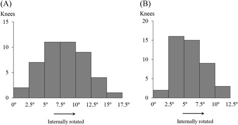
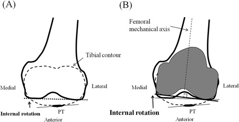
Results: The distal surface of femoral condyles was internally rotated relative to the tibial attachment of the patellar tendon and the equatorial line of the patella in all the knees (8.2° ± 3.5° and 5.8° ± 2.5°, respectively), not parallel.
Conclusions: Distal femoral condyle is internally rotated to the patellar tendon at 90° of flexion in normal knees. When the symmetric femoral component is aligned perpendicular to the femoral mechanical axis, the patellar tendon would be possibly more twisted than the condition in normal knees, and the deviation of the PF contact force on the patellar component might be caused. The configuration and alignment of the distal condyle of the femoral component can affect the PF joint congruity in knee flexion. In this respect, our results provide important information in considering designs and alignment in the distal femur of TKA and the PF joint congruity in knee flexion.
Figures
References
-
- Parker DA, Dunbar MJ, Rorabeck CH. Extensor mechanism failure associated with total knee arthroplasty: prevention and management. J Am Acad Orthop Surg. 2003;11:238–47. - PubMed
-
- Parratte S, Pagnano MW. Instability after total knee arthroplasty. Instr Course Lect. 2008;57:295–304. - PubMed
-
- Sharma A, Komistek RD, Scuderi GR, Cates HE., Jr High-flexion TKA designs: what are their in vivo contact mechanics? Clin Orthop Relat Res. 2007;464:117–26. - PubMed
Publication types
MeSH terms
LinkOut - more resources
Full Text Sources
Other Literature Sources
