

doi: 10.1038/cr.2015.49.
Epub 2015 Apr 24.
Paternally contributed centrioles exhibit exceptional persistence in C. elegans embryos
Affiliations
- PMID: 25906994
- PMCID: PMC4423087
- DOI: 10.1038/cr.2015.49
Item in Clipboard
Paternally contributed centrioles exhibit exceptional persistence in C. elegans embryos
Cell Res.
2015 May.
No abstract available
Figures
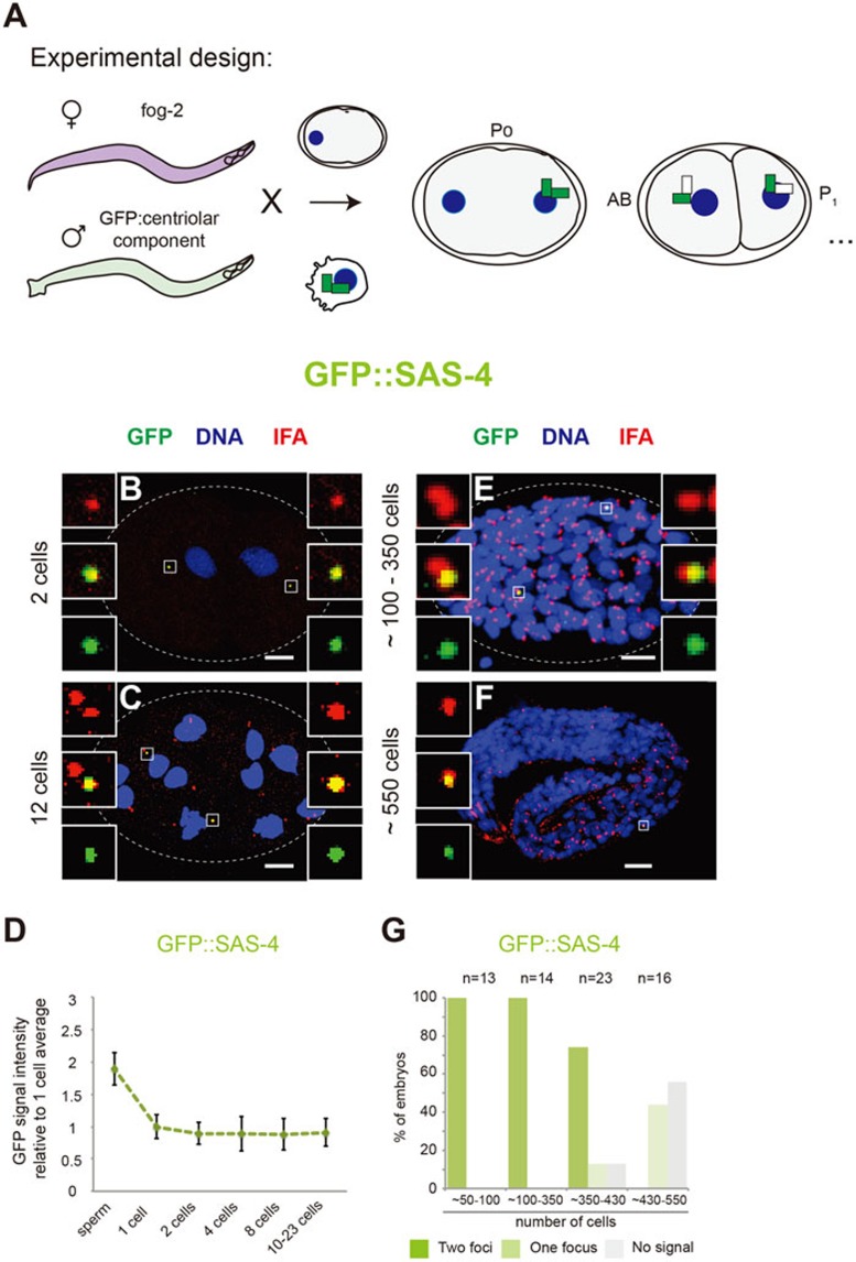
Paternally contributed centriolar components persist throughout C. elegans embryogenesis. (A) Marked mating experimental strategy to track paternally contributed centriolar components during C. elegans embryogenesis. Feminized fog-2 animals were crossed with males expressing given GFP-tagged centriolar components in the germline; the zygotes generated thereby inherited paternally contributed GFP-positive centrioles. Thereafter, centrioles formed in the embryo were assembled using the maternally contributed cytoplasmic pool of proteins, which is GFP negative. (B, C, E, F)
C. elegans embryos at the indicated stages stemming from marked mating experiments with GFP::SAS-4 males. Embryos were stained with antibodies against GFP (green) and the pan-centriolar marker IFA (red), as well as with Hoechst to reveal DNA (blue). Scale bar, 5 μm. Insets are 5-time magnified views of centrioles. Note that as a result of unspecific immunostaining, small background green foci not colocalizing with IFA can be observed in some images; such foci were not taken into consideration for analysis. (D) Quantification of the GFP signal of paternally contributed centrioles at the indicated stages of early development for GFP::SAS-4 by immunofluorescence analysis. Signal intensity is expressed relative to the average signal of the 1-cell stage embryos. The approximately two-fold intensity observed in sperm compared to 1-cell stage embryos reflects the fact that sperm cells carry two GFP-tagged centrioles. Number of centrioles analyzed: sperm, n = 417; 1 cell, n = 20; 2 cells, n = 14; 4 cells, n = 16; 8 cells, n = 16; 10-23 cells, n = 20. (G) Percentage of embryos with 1, 2 or no GFP-positive paternally contributed centrioles from marked mating experiments with GFP::SAS-4 at the indicated stages of embryogenesis.
References
Publication types
MeSH terms
Substances
LinkOut - more resources
Full Text Sources
Other Literature Sources
