. 2015 Jun:54:168-76.
doi: 10.1016/j.biomaterials.2015.03.020. Epub 2015 Apr 7.

Fabrication and characterization of medical grade polyurethane composite catheters for near-infrared imaging

Affiliations

Fabrication and characterization of medical grade polyurethane composite catheters for near-infrared imaging

André T Stevenson Jr et al. Biomaterials. 2015 Jun.

Abstract

Peripherally inserted central catheters (PICCs) are hollow polymeric tubes that transport nutrients, blood and medications to neonates. To determine proper PICC placement, frequent X-ray imaging of neonates is performed. Because X-rays pose severe health risks to neonates, safer alternatives are needed. We hypothesize that near infrared (NIR) polymer composites can be fabricated into catheters by incorporating a fluorescent dye (IRDye 800CW) and visualized using NIR imaging. To fabricate catheters, polymer and dye are dry mixed and pressed, sectioned, and extruded to produce hollow tubes. We analyzed surface roughness, stiffness, dye retention, NIR contrast intensity, and biocompatibility. The extrusion process did not significantly alter the mechanical properties of the polymer composites. Over a period of 23 days, only 6.35 ± 5.08% dye leached out of catheters. The addition of 0.025 wt% dye resulted in a 14-fold contrast enhancement producing clear PICC images at 1 cm under a tissue equivalent. The addition of IRDye 800CW did not alter the biocompatibility of the polymer and did not increase adhesion of cells to the surface. We successfully demonstrated that catheters can be imaged without the use of harmful radiation and still maintain the same properties as the unaltered medical grade equivalent.

Keywords: Catheter; Cell adhesion; Fluorescence; Mechanical testing; Polyurethane.


Figures

Fig. 1
Fig. 1
Schematic of the fabrication process for Composite Catheters.
Fig. 2
Fig. 2
Optical images (A-D) and SEM micrographs (E-P) of polymer samples. Hospital TPU (A) appears perfectly round and smooth. The extruded samples (B, C, D) appear smooth and optically transparent similar to the Hospital TPU with the composite samples being nearly indistinguishable from their unmodified counterparts. SEM micrographs consist of cross sectional view (E,F,G,H), top view (I,J,K,L), and roughness profiles (M,N,O,P). Collectively, the extruded samples have large diameters and thicknesses compared to the Hospital TPU due to the extruder die design (Table 1). Plain TPU (F), TPU Composite (G) and Leached TPU Composite (H) have irregular cross sectional slices due to swelling and sample collection during extrusion. Top view and roughness images between all samples appear similar. Optical image scale bar = 7.5 mm, cross sectional and top view scale bar = 200 μm, roughness image scale bar = 600 nm.
Fig. 3
Fig. 3
Mechanical Properties of Catheters. Elastic modulus (Table 3) was determined by the slope of the linear region between 0 to 10% strain. The UTS (Table 3) was determined to be the point at which the samples fractured. Error bars represent standard deviations for every 100 data points per sample (n=3).
Fig. 4
Fig. 4
Retention analysis of IR Dye 800 CW within TPU matrix. 6.35 ± 5.08% of the IR Dye 800 CW was released from the polymer over 23 days. Inset is the same data with axis adjusted for clarity. The majority of the dye released as a burst within the first five days (5.40%) followed my minimal leaching throughout the duration of the study. Error bars represents standard deviations (n=8).
Fig. 5
Fig. 5
Fluorescence intensity scans of Plain TPU, TPU Composite, and Leached TPU Composite. Samples were imaged at an excitation wavelength of 778 nm. 0, 1, 2, 3 cm correspond to the imaging depth or the thickness of Superflab covering the samples that the imaging probe penetrated.
Fig. 6
Fig. 6
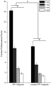
Contrast Enhancement intensity factor of TPU Composites. The fluorescence intensity decreases as a function of depth, though signal is still observed at 3 cm. All values are statistically different within the non-leached and leached samples. All values are statistically different between the non-leached and leached samples except for at 3 cm. Error bars represent standard deviation (n=4). Asterisk (*) represents statistically significant data (p<0.05).
Fig. 7
Fig. 7
Biocompatibility of thin films with HUVECs. Cells are stained with Calcein AM (green) to indicate viable cells and propidium iodide (red) to signify dead cells. Incubation of cells with Plain TPU, TPU Composite and 0.025 wt% IR Dye resulted in a non-significant difference in viability compared to the control. No apparent changes in morphology or proliferation were observed due to the polymer composite, polymer or IRDye 800CW.
Fig. 8
Fig. 8
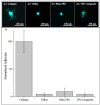
Adhesion studies of HUVECs seeded directly on top of substrates. (A1-A4) Typical morphologies of HUVECs stained with Calcein AM (green) on respective substrates. Collagen preferentially adhered to Collagen I with substantial cell spreading. Cells weakly attached to Teflon, Plain TPU, and TPU Composite as evidenced by the round shape with no extended lamellipodia. (B) Normalized numbers of cells adhered to substrates over a 60 minute time period. Cells had a high affinity for collagen I, one of the major proteins found in HUVECs native microenvironment. Minimal adherence occurred for Teflon, Plain TPU, and TPU Composite. Adherences of cells for the three films (Teflon, Plain TPU, TPU Composite) are statistically different from the collagen with no difference observed between the three polymer films.

References

    1. McCay AS, Elliott EC, Walden M. PICC Placement in the Neonate. New England Journal of Medicine. 2014;370(11) - PubMed
    1. Vo JN, Hoffer FA, Shaw DW. Techniques in vascular and interventional radiology: pediatric central venous access. Techniques in vascular and interventional radiology. 2010;13(4):250–7. - PubMed
    1. Hogan MJ. Neonatal vascular catheters and their complications. Radiologic Clinics of North America. 1999;37(6):1109–25. - PubMed
    1. Sharpe EL. Repositioning techniques for malpositioned neonatal peripherally inserted central catheters. Advances in Neonatal Care. 2010;10(3):129–32. - PubMed
    1. Patricia Catudal J, Sharpe EL. The Wandering Ways of a PICC Line: Case Report of a Malpositioned Peripherally Inserted Central Catheter (PICC) and Correction. Journal of the Association for Vascular Access. 2011;16(4):218–20.

Publication types