. 2015 Apr 24:16:26.
doi: 10.1186/s12868-015-0166-2.

Presynaptic BK channel localization is dependent on the hierarchical organization of alpha-catulin and dystrobrevin and fine-tuned by CaV2 calcium channels

Affiliations

Presynaptic BK channel localization is dependent on the hierarchical organization of alpha-catulin and dystrobrevin and fine-tuned by CaV2 calcium channels

Kelly H Oh et al. BMC Neurosci. .

Abstract

Background: Large conductance, calcium-activated BK channels regulate many important physiological processes, including smooth muscle excitation, hormone release and synaptic transmission. The biological roles of these channels hinge on their unique ability to respond synergistically to both voltage and cytosolic calcium elevations. Because calcium influx is meticulously regulated both spatially and temporally, the localization of BK channels near calcium channels is critical for their proper function. However, the mechanism underlying BK channel localization near calcium channels is not fully understood.

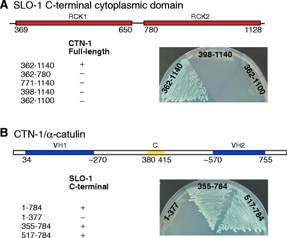
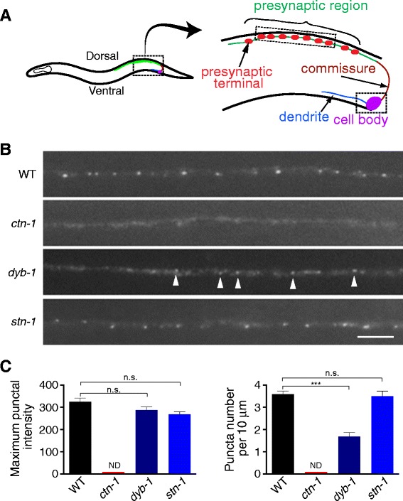
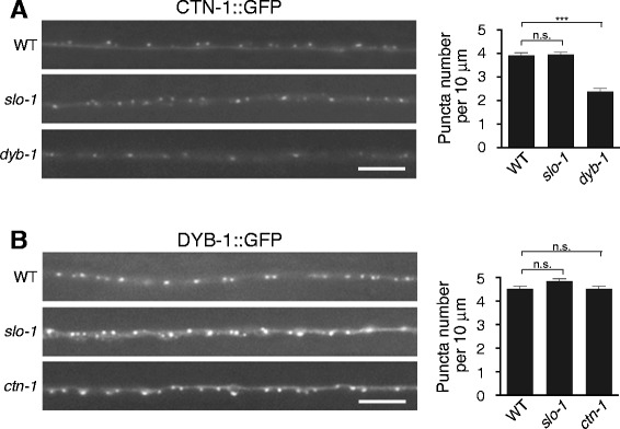
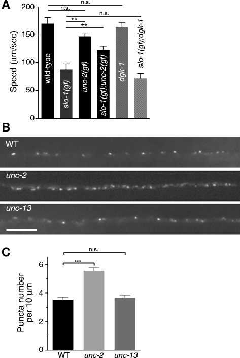
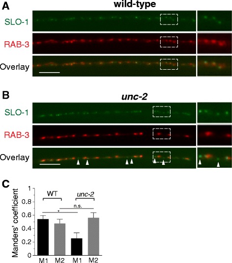
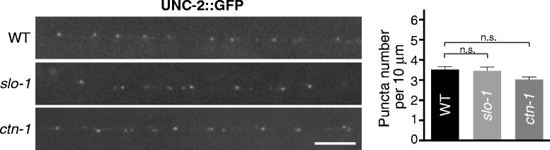
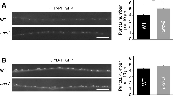
Results: We show here that in C. elegans the localization of SLO-1/BK channels to presynaptic terminals, where UNC-2/CaV2 calcium channels regulate neurotransmitter release, is controlled by the hierarchical organization of CTN-1/α-catulin and DYB-1/dystrobrevin, two proteins that interact with cortical cytoskeletal proteins. CTN-1 organizes a macromolecular SLO-1 channel complex at presynaptic terminals by direct physical interaction. DYB-1 contributes to the maintenance or stabilization of the complex at presynaptic terminals by interacting with CTN-1. We also show that SLO-1 channels are functionally coupled with UNC-2 calcium channels, and that normal localization of SLO-1 to presynaptic terminals requires UNC-2. In the absence of UNC-2, SLO-1 clusters lose the localization specificity, thus accumulating inside and outside of presynaptic terminals. Moreover, CTN-1 is also similarly localized in unc-2 mutants, consistent with the direct interaction between CTN-1 and SLO-1. However, localization of UNC-2 at the presynaptic terminals is not dependent on either CTN-1 or SLO-1. Taken together, our data strongly suggest that the absence of UNC-2 indirectly influences SLO-1 localization via the reorganization of cytoskeletal proteins.

Conclusion: CTN-1 and DYB-1, which interact with cortical cytoskeletal proteins, are required for the presynaptic punctate localization of SLO-1 in a hierarchical manner. In addition, UNC-2 calcium channels indirectly control the fidelity of SLO-1 puncta localization at presynaptic terminals. We suggest that the absence of UNC-2 leads to the reorganization of the cytoskeletal structure that includes CTN-1, which in turn influences SLO-1 puncta localization.


Figures

Figure 1
Figure 1
Yeast two-hybrid assays reveal the interaction between CTN-1/α-catulin and the two RCK domains of SLO-1/BK. (A) Both the RCK1 and RCK2 domains of SLO-1 are necessary for the interaction with CTN-1. The indicated portions of the SLO-1 C-terminal region were fused in frame to the GAL4 activation domain and tested for interaction with CTN-1 tagged with the GAL4 DNA-binding domain. Images obtained from different plates cultured under identical conditions are stitched together for the simplicity of presentation. (B) The C-terminal region of CTN-1 that encompasses the VH2 domain interacts with SLO-1. The indicated portions of CTN-1 were fused in frame to the GAL4-DNA binding domain and tested for interaction with the SLO-1 C-terminal region tagged with the GAL4 activation domain. VH1, vinculin homology domain 1; C, coiled-coil domain; VH2, vinculin homology domain 2.
Figure 2
Figure 2
SLO-1/BK localization at presynaptic terminals is controlled by CTN-1/α-catulin and DYB-1/dystrobrevin, but not by STN-1/syntrophin. (A) Schematic representation of the anatomical structure of a DA neuron. The DB neurons have an axon and dendrites that extend in the other direction. The unc-129 promoter (~2. 4 kb) drives expression in a subset of DA and DB cholinergic motor neurons. The axon processes with neuromuscular and VD synapses (green) are presynaptic regions, and individual synaptic areas (red) are presynaptic terminals. (B) Representative images of SLO-1::GFP in the axon terminals of DA and DB neurons in wild-type, ctn-1, dyb-1 and stn-1 mutant animals. Arrowheads highlight SLO-1::GFP puncta. The scale bar represents 5 μm. (C) Quantification of SLO-1::GFP puncta in wild-type and mutant animals. The maximum intensity of puncta and the number of puncta within a 10 μm distance were calculated using dotGUI (see Methods). For all the panels, data are presented as the mean ± SEM. *** and n.s. indicate a statically significant difference (multiplicity adjusted p < 0.001) and no significant difference between indicated groups, respectively (One-way ANOVA Dunett’s multiple comparison. wild-type, n = 11; dyb-1, n = 12; stn-1; n = 5). ND, not determined.
Figure 3
Figure 3
Presynaptic localization of CTN-1/α-catulin and DYB-1/dystrobrevin (A) Presynaptic localization of CTN-1/α-catulin is partially dependent on DYB-1/dystrobrevin. The integrated array cimIs8, which drives GFP-tagged CTN (GFP::CTN-1) expression in a subset of DA and DB neurons, was crossed to slo-1 or dyb-1 mutants. The number of puncta within 10 μm in each data point was presented as the mean ± SEM and analyzed by one-way ANOVA Dunett’s multiple comparison (wild-type, n = 20; slo-1, n = 29; dyb-1, n = 20. ***p < 0.001; n.s. p > 0.05). (B) Presynaptic localization of DYB-1/dystrobrevin is independent of CTN-1/α-catulin. The integrated array cimIs15, which drives GFP-tagged DYB (GFP::DYB-1) expression in a subset of DA and DB neurons, was crossed to slo-1 or ctn-1 mutants. The number of puncta within 10 μm in each data point was presented as the mean ± SEM (one-way ANOVA Dunett’s multiple comparison, wild-type, n = 34; slo-1, n = 31; ctn-1, n = 30; n.s. p > 0.05). The scale bar represents 5 μm.
Figure 4
Figure 4
Genetic interaction of SLO-1/BK and UNC-2/CaV2 channels. (A) SLO-1/BK functionally interacts with UNC-2/CaV2 channels for locomotory behavior. A gain-of-function unc-2(zf35) mutation suppresses the sluggish movement of slo-1(gf) mutants, whereas dgk-1 mutation, which causes a hyperactive phenotype, does not. The data are presented as the mean ± SEM and analyzed by one-way ANOVA with Bonferroni’s post Hoc test (**p < 0.01, n.s., not significant). (B and C) A loss-of-function unc-2 mutation increases the SLO-1 punctal density in the presynaptic region. The number of SLO-1 puncta in a given length of the axonal terminal (i.e., decreased punctal distance) is higher in unc-2 mutants than wild-type or unc-13 animals. The quantification of SLO-1 puncta number was performed in cimIs10 animals whose genetic background is wild-type, unc-2 or unc-13 animals. The data are presented as the mean ± SEM and analyzed by two-tailed Student t-test (wild-type, n = 14; unc-2, n = 11; unc-13, n = 12 ***p < 0.001, n.s., not significant). Scale bar, 5 μm.
Figure 5
Figure 5
The organization of SLO-1/BK at the presynaptic terminals is defective in unc-2 mutants. (A, B) An integrated SLO-1::GFP array cimIs10 was crossed with wild-type (A) or unc-2 (B) animals carrying the integrated RAB-3::mCherry array cimIs14. Arrowheads indicate SLO-1 puncta that are not co-localized with clusters of synaptic vesicles shown by RAB-3 puncta. The scale bars represent 5 μm. (C) Co-localization between SLO-1::GFP and RAB-3::mCherry in wild-type and unc-2 animals was analyzed with JACoP, an ImageJ plugin [44]. Manders’ coefficient M1 is defined as the ratio of the summed intensity of GFP pixels overlapped with mCherry to total GFP intensity, whereas M2 is conversely defined. M1 coefficients in wild-type (n = 5) and unc-2 (n = 4) animals are significantly different, whereas M2 coefficients in both animals are not different (Student t-test, n.s., p = 0.34; *p = 0.013). The data represent mean ± SEM.
Figure 6
Figure 6
UNC-2/CaV2 localization is independent of SLO-1/BK localization. slo-1 or ctn-1 null mutants do not have any obvious defect in UNC-2 localization. Left panels: representative image. Right panels: quantification of the puncta number. Data were analyzed by one-way ANOVA with Dunnett’s multiple comparisons test (wt vs. slo-1: P = 0.94; wt vs. ctn-1: P = 0.07).
Figure 7
Figure 7
The disruption of SLO-1/BK localization in unc-2 mutants is caused by altered CTN-1/α-catulin localization. (A) CTN-1 localization in wild-type and unc-2 mutant animals. The number of GFP::CTN-1 puncta was increased in unc-2 mutants compared with wild-type animals. The data are presented as the mean ± SEM and analyzed by two-tailed Student’s t-test (wild-type, n = 28; unc-2, n = 25; ***p < 0.001). (B) DYB-1 localization in wild-type and unc-2 mutant animals. The number of GFP::DYB-1 puncta was slightly increased in unc-2 mutants compared with wild-type animals. The data are presented as mean ± SEM and analyzed by two-tailed Student’s t-test (wild-type, n = 46; unc-2, n = 18; *p < 0.05). The scale bar represents 5 μm.

References

    1. Clapham DE. Calcium signaling. Cell. 2007;131(6):1047–58. doi: 10.1016/j.cell.2007.11.028. - DOI - PubMed
    1. Simms BA, Zamponi GW. Neuronal voltage-gated calcium channels: structure, function, and dysfunction. Neuron. 2014;82(1):24–45. doi: 10.1016/j.neuron.2014.03.016. - DOI - PubMed
    1. Fakler B, Adelman JP. Control of K(Ca) channels by calcium nano/microdomains. Neuron. 2008;59(6):873–81. doi: 10.1016/j.neuron.2008.09.001. - DOI - PubMed
    1. Yuan P, Leonetti MD, Pico AR, Hsiung Y, MacKinnon R. Structure of the human BK channel Ca2+−activation apparatus at 3.0 A resolution. Science. 2010;329(5988):182–6. doi: 10.1126/science.1190414. - DOI - PMC - PubMed
    1. Horrigan FT, Aldrich RW. Allosteric voltage gating of potassium channels II. Mslo channel gating charge movement in the absence of Ca(2+) J Gen Physiol. 1999;114(2):305–36. doi: 10.1085/jgp.114.2.305. - DOI - PMC - PubMed

Publication types

MeSH terms

Substances

LinkOut - more resources