Review
. 2015 Sep;39(5):649-69.
doi: 10.1093/femsre/fuv015. Epub 2015 Apr 22.

Giving structure to the biofilm matrix: an overview of individual strategies and emerging common themes

Affiliations
Review

Giving structure to the biofilm matrix: an overview of individual strategies and emerging common themes

Laura Hobley et al. FEMS Microbiol Rev. 2015 Sep.

Abstract

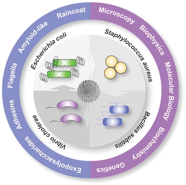
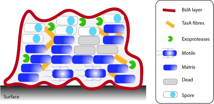
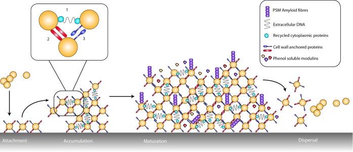
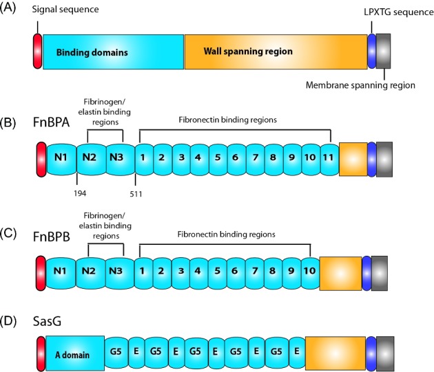
Biofilms are communities of microbial cells that underpin diverse processes including sewage bioremediation, plant growth promotion, chronic infections and industrial biofouling. The cells resident in the biofilm are encased within a self-produced exopolymeric matrix that commonly comprises lipids, proteins that frequently exhibit amyloid-like properties, eDNA and exopolysaccharides. This matrix fulfils a variety of functions for the community, from providing structural rigidity and protection from the external environment to controlling gene regulation and nutrient adsorption. Critical to the development of novel strategies to control biofilm infections, or the capability to capitalize on the power of biofilm formation for industrial and biotechnological uses, is an in-depth knowledge of the biofilm matrix. This is with respect to the structure of the individual components, the nature of the interactions between the molecules and the three-dimensional spatial organization. We highlight recent advances in the understanding of the structural and functional role that carbohydrates and proteins play within the biofilm matrix to provide three-dimensional architectural integrity and functionality to the biofilm community. We highlight, where relevant, experimental techniques that are allowing the boundaries of our understanding of the biofilm matrix to be extended using Escherichia coli, Staphylococcus aureus, Vibrio cholerae, and Bacillus subtilis as exemplars.

Keywords: Bacillus subtilis; Escherichia coli; Staphylococcus aureus; Vibrio cholerae; amyloid fibres; biofilm matrix assembly; biophysics; hydrophobin.


Figures

Graphical Abstract Figure.
Examining the structure and function of the biofilm extracellular matrix.
Figure 1.
Figure 1.
β-sheet-rich fibre formation by E. coli, B. subtilis and S. aureus. (A) Curli are amyloidous protein fibres assembled on the surface of E. coli cells within the nutrient-depleted zones of a biofilm and provide structural integrity. The curli fibre subunit CsgA is exported across the outer membrane through the CsgG translocator channel. Once outside the cell, CsgA interacts with the CsgB nucleator protein, and polymerizes into amyloidous fibres that extend away from the cell. The accessory proteins CsgC and CsgE regulate export by CsgG and CsgF is required for nucleation of CsgA by CsgB. (B) TasA-amyloid-like β-sheet-rich fibres protrude from the cell wall and are required for biofilm formation. Biogenesis requires the products of the tapA-sipW-tasA operon. Each protein is made in the cytoplasm and transported across the membrane by the Sec export system. SipW functions as a dedicated signal peptidase to cleave the signal peptide from TasA and TapA. TapA is required for anchoring the fibres to the cell wall and forms a minor component in the β-sheet-rich fibres. TasA is the major component in the fibres. (C) PSM β-rich fibres are found elaborated on the surface of S. aureus. The PSM transporter (PMT) is an ATP-dependent ABC transporter. It is composed of two transmembrane proteins (PmtB and D), coupled with the ATPases PmtA and PmtC. PSMs are known to function in the monomeric state where their surfactant activity has cytolytic activity. The formation of the fibre form is proposed to be a mechanism to inactivate the monomers until they are required again.
Figure 2.
Figure 2.
Escherichia coli biofilm structure. After prolonged incubation, E. coli forms complex colony type biofilms on agar plates, where the three major components of the biofilm matrix have been shown to be curli fibres, cellulose and flagella filaments. Left: the E. coli complex colony contains both concentric rings and axial wrinkles, the rings are dependent on the production of both curli and flagella and the axial wrinkles additionally require the production of cellulose. The colony can be divided into three zones (I) the outer edge, (II) the middle zone and (III) the inner region. Right: a cross-section of the colony shows the different cell types co-existing within the biofilm, and their location within the different biofilm regions. Dividing, flagellated cells are found within the outer edge, whilst in the middle zone and the inner region two distinct cell types are found. Near the agar surface post-exponential, rod-shaped cells are found encased in a mesh of flagella filaments, whilst in the upper levels of the colony stationary phase, ovoid cells are found, these are surrounded in a dense mesh of curli fibres and cellulose.
Figure 3.
Figure 3.
Structure and deployment of V. cholerae biofilm matrix proteins. (A) Bioinformatics analysis of RmbC (accession number Q9KTH2), RmbA (accession number Q9KTH4) and Bap1 (accession number Q9KQW0) was performed de novo using a combination of SMART (Schultz et al. ; Letunic, Doerks and Bork 2014), InterPro (Hunter et al. 2012) and BLAST (Johnson et al. 2008) to identify conserved domains, SignalP (Petersen et al. 2011) to designate signal sequence peptide cleavage sites and where required further information was revealed using WU-BLAST analysis (http://www.ebi.ac.uk/Tools/sss/wublast/). For Bap1, it should be noted that the EF-hand domain has a low confidence score of 7.00e-03 and for RmbC the integrin α-N-terminal domain had a confidence value of 5.00e-04 so the presence of these protein domains should be interpreted with caution. The domains and proteins are drawn approximately to scale. Parts B and C are reproduced, with permission, from Berk et al. (2012) Science along with the corresponding legend. Images are pseudo-coloured blue (cells), grey (RbmA), red (RbmC) and green (Bap1). RbmA localizes around and within cell clusters. RbmC and Bap1 encase cell clusters. Cells were counterstained with DAPI. Scale bars, 3 μm. (B) 3D biofilm architecture. (C) Enlargement of the boxed region in (B). Red arrow indicates one cell cluster. Red signal now rendered partially transparent to allow visualization of cells within an RbmC-containing cluster.
Figure 4.
Figure 4.
Bacillus subtilis biofilm formation. Biofilm formation by B. subtilis culminates in the formation of a structured highly hydrophobic sessile community. The isogenic population differentiates to divide tasks within the community. For a detailed review of this process refer to (Cairns, Hobley and Stanley-Wall ; Mielich-Suss and Lopez 2014).
Figure 5.
Figure 5.
Staphylococcus aureus biofilm formation. Attachment of S. aureus to a surface is mediated by CWA proteins. Cell-to-cell interactions occur during accumulation phase and can be mediated by several factors. The magnified region shows this in more detail: (1) extracellular DNA linking recycled cytoplasmic proteins; (2) CWA proteins binding adjacent cell surfaces; (3) Homophilic interactions between CWA proteins. PSMs form amyloid-like fibres visible at the surface of the biofilm. They also act in the formation of channels within the biofilm to allow nutrient access, while their surfactant properties aid the dispersal phase. The different stages of biofilm formation are detailed from left to right across the diagram.
Figure 6.
Figure 6.
Structure of the CWA proteins. All CWA proteins contain a Sec-dependent secretory signal sequence, a C-terminal LPXTG sortase motif, a hydrophobic domain and finally a chain of positively charged residues at the end of the C-terminus. (A) The schematic demonstrates the typical domain structure within the MSCRAMM family of CWAs. At the N-terminus, a Sec-dependent signal sequence followed by a variable number of binding domains that begin with an N-terminal A domain (inclusive of N subdomains in the case of FnBPs (B and C) and ClfB). These are followed by a wall-spanning region, the LPXTG motif and finally a membrane-spanning region. (D) In SasG, the binding domain is subdivided into the N-terminal A domain and a varying number of G5/E repeats.

References

    1. Abraham NM, Jefferson KK. Staphylococcus aureus clumping factor B mediates biofilm formation in the absence of calcium. Microbiology. 2012;158:1504–12. - PMC - PubMed
    1. Absalon C, Van Dellen K, Watnick PI. A communal bacterial adhesin anchors biofilm and bystander cells to surfaces. PLoS Pathog. 2011;7:e1002210. - PMC - PubMed
    1. Altindis E, Fu Y, Mekalanos JJ. Proteomic analysis of Vibrio cholerae outer membrane vesicles. P Natl Acad Sci USA. 2014;111:E1548–56. - PMC - PubMed
    1. Andersson EK, Bengtsson C, Evans ML, et al. Modulation of curli assembly and pellicle biofilm formation by chemical and protein chaperones. Chem Biol. 2013;20:1245–54. - PMC - PubMed
    1. Asally M, Kittisopikul M, Rue P, et al. Localized cell death focuses mechanical forces during 3D patterning in a biofilm. P Natl Acad Sci USA. 2012;109:18891–6. - PMC - PubMed

Publication types