Review
. 2015 May;16(5):299-309.
doi: 10.1038/nrm3980.

Somatic stem cell heterogeneity: diversity in the blood, skin and intestinal stem cell compartments

Affiliations
Review

Somatic stem cell heterogeneity: diversity in the blood, skin and intestinal stem cell compartments

Margaret A Goodell et al. Nat Rev Mol Cell Biol. 2015 May.

Abstract

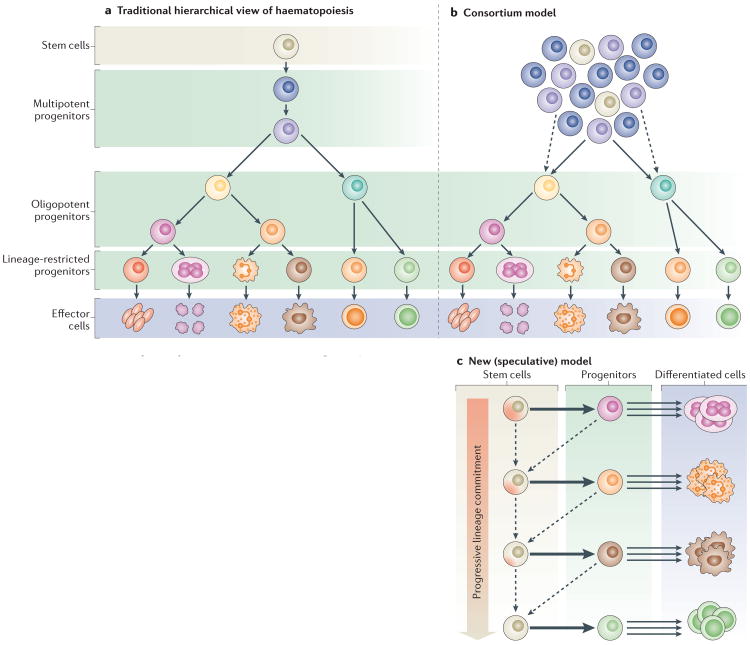
Somatic stem cells replenish many tissues throughout life to repair damage and to maintain tissue homeostasis. Stem cell function is frequently described as following a hierarchical model in which a single master cell undergoes self-renewal and differentiation into multiple cell types and is responsible for most regenerative activity. However, recent data from studies on blood, skin and intestinal epithelium all point to the concomitant action of multiple types of stem cells with distinct everyday roles. Under stress conditions such as acute injury, the surprising developmental flexibility of these stem cells enables them to adapt to diverse roles and to acquire different regeneration capabilities. This paradigm shift raises many new questions about the developmental origins, inter-relationships and molecular regulation of these multiple stem cell types.


Figures

Figure 1
Figure 1. Stem cell models for the haematopoietic system
a | The traditional hierarchical view of haematopoiesis is that there is one type of stem cell that has the capacity to give rise to lineage-restricted progenitors that differentiate into all the cell types of the blood with equivalent propensity. b | In the consortium model, a pool of stem cells with slightly different properties regenerates the system continuously through progenitors that are increasingly restricted in their potential. c | In a new speculative model, stem cells are rare reserve cells that occasionally generate lineage-restricted progenitors. These stem cells have different lineage biases and give rise to specific progenitors. Existing data suggest that the most primitive stem cells are primed towards the megakaryocyte lineage. These stem cells give rise to progenitors that are largely restricted to specific fate choices, and these progenitors are the main drivers of haematopoiesis, generating massive numbers of differentiated cells over a long period of time. During extreme stress (such as major injury or transplantation), the progenitors may revert (dashed arrows pointing left) to a stem-like state while retaining some of their lineage preferences. This model is consistent with the reported existence of megakaryocyte-biased stem cells (cells at the top of the progenitor hierarchy) and lymphoid-primed multipotent progenitors (one-step-down stem-like cells that lack megakaryocyte differentiation potential) as well as with the increasing differentiation bias observed with age. Indeed, it has been shown that the progenitors lose their developmental flexibility during ageing. Models that are hybrids of the three that are outlined in this figure can also be envisioned.
Figure 2
Figure 2. Heterogeneity of skin epithelial stem cells
The resting-phase hair follice(HF) contains several compartments (indicated by different colours), which are defined by cells that express distinct molecular markers: stem cell antigen 1 (Sca1; also known as Ly6a), leucine-rich repeats and immunoglobulin-like domains 1 (Lrig1), placenta-expressed transcript 1 (Plet1), leucine-rich repeat-containing G protein-coupled receptor 6 (Lgr6), Blimp1, Sox9, transcription factor 3 (Tcf3), Tcf4, LIM homeobox 2 (Lhx2), keratin 15 (K15), K19, CD34, nuclear factor of activated T cells, cytoplasmic calcineurin-dependent 1 (Nfatc1) and Gli1. The compartments above the HF bulge include the infundibulum, which is contiguous with the interfollicular epidermis (IFE) in the uppermost portion of the HF; the isthmus, which is located directly below the infundibulum; and the junctional zone, which is part of the upper isthmus and lies next to the sebaceous gland (SG). These compartments contain their own stem cells that maintain their homeostasis. The bulge itself contains HF stem cells, whereas the hair germ (HG), which is located directly below the bulge, comprises progenitor cells. The HG is marked by high levels of cadherin 3 (Cdh3) expression and is situated on top of the dermal papilla, which is a cluster of specialized mesenchymal cells that are required for HF regeneration. HFs comprise a heterogeneous population of stem cells, which express different markers as indicated. As listed in the table, lineage tracing using inducible Cre recombinases under the control of specific promoters, which are expressed in distinct cell populations, shows the restricted lineage potential of the heterogeneous populations during normal homeostasis as well as an expanded potential in response to injury. ND, not done.
Figure 3
Figure 3. Overview of the intestinal stem cell system
a | Intestinal progenitor cells are organized in crypts of Lieberkühn. Intestinal stem cells (ISCs) reside near the crypt base and produce daughter cells (transient-amplifying (TA) cells), which proliferate in the mid-crypt and terminally differentiate near the crypt opening to produce the diverse range of intestinal epithelial cell types. Crypt base columnar (CBC) stem cells are interdigitated with Paneth cells at the crypt base. The differentiated cells that line the villus include absorptive enterocytes, goblet cells and enteroendocrine cells. Quiescent ISCs (qISCs; also known as +4 stem cells) are thought to reside just above the zone that contains Paneth and CBC cells. b | CBC cells compete for niche support and space at the base of the crypt, which confers a competitive advantage on the lower (tier 1) CBC cells. Tier 2 CBC cells differentiate as they leave the niche, using contact-dependent Notch signalling to determine their cell fate. Contact with ligand-expressing secretory cells (goblet, enteroendocrine or Paneth cells) activates Notch and directs cells into the absorptive lineage, which gives rise to enterocytes. Contact that is limited to enterocytes or their progenitors results in expression of the transcription factor atonal homologue 1 (ATOH1) and in secretory lineage commitment. Secretory progenitor cells subsequently differentiate into goblet, Paneth or enteroendocrine cells. Extreme injury that causes depletion of the CBC cell pool can induce reversion of committed progenitor cells to a stem cell state (indicated by the dashed arrow). qISCs are thought to give rise to CBC cells following injury. It remains unclear whether a dedicated pool of qISCs exists or whether these cells are transient progenitor cells under homeostatic conditions.

References

    1. Pappenheim A. Virchows Arch Pathol Anat Physiol Klin Med. Vol. 145. in German: 1896. Ueber Entwickelung und Ausbildung der Erythroblasten; pp. 587–643.
    1. Ramalho-Santos M, Willenbring H. On the origin of the term “stem cell”. Cell Stem Cell. 2007;1:35–38. - PubMed
    1. Becker AJ, McCulloch EA, Till JE. Cytological demonstration of the clonal nature of spleen colonies derived from transplanted mouse marrow cells. Nature. 1963;197:452–454. - PubMed
    1. Lemischka IR, Raulet DH, Mulligan RC. Developmental potential and dynamic behavior of hematopoietic stem cells. Cell. 1986;45:917–927. - PubMed
    1. Keller G, Snodgrass R. Life span of multipotential hematopoietic stem cells in vivo. J Exp Med. 1990;171:1407–1418. - PMC - PubMed

Publication types