. 2015 Apr 24;17(1):57.
doi: 10.1186/s13058-015-0569-0.

Adipose cells promote resistance of breast cancer cells to trastuzumab-mediated antibody-dependent cellular cytotoxicity

Affiliations

Adipose cells promote resistance of breast cancer cells to trastuzumab-mediated antibody-dependent cellular cytotoxicity

Minh Ngoc Duong et al. Breast Cancer Res. .

Abstract

Introduction: Trastuzumab has been used in the treatment of human epidermal growth factor receptor 2 (HER2)-expressing breast cancer, but its efficacy is limited by de novo or acquired resistance. Although many mechanisms have been proposed to explain resistance to trastuzumab, little is known concerning the role of the tumor microenvironment. Given the importance of antibody-dependent cellular cytotoxicity (ADCC) in the antitumor effect of trastuzumab and the abundance of adipose tissue in the breast, we investigated the impact of adipocytes on ADCC.

Methods: We set up a coculture system to study the effect of adipocytes on ADCC in vitro. The results were validated in vivo in a mouse xenograft model.

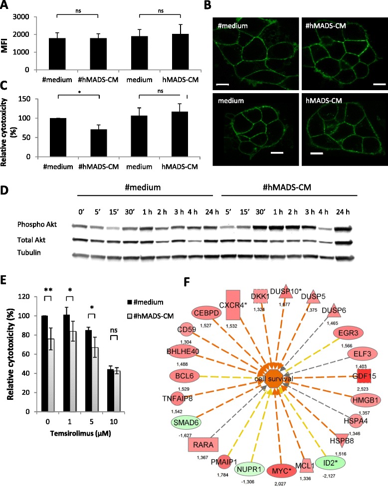
Results: We found that adipocytes, as well as preadipocytes, inhibited trastuzumab-mediated ADCC in HER2-expressing breast cancer cells via the secretion of soluble factors. The inhibition of ADCC was not due to titration or degradation of the antibody. We found that adipose cells decreased the secretion of interferon-γ by natural killer cells, but did not alter natural killer cells' cytotoxicity. Preincubation of breast cancer cells with the conditioned medium derived from adipocytes reduced the sensitivity of cancer cells to ADCC. Using a transcriptomic approach, we found that cancer cells undergo major modifications when exposed to adipocyte-conditioned medium. Importantly, breast tumors grafted next to lipomas displayed resistance to trastuzumab in mouse xenograft models.

Conclusions: Collectively, our findings underline the importance of adipose tissue in the resistance to trastuzumab and suggest that approaches targeting the adipocyte-cancer cell crosstalk may help sensitize cancer cells to trastuzumab-based therapy.


Figures

Figure 1
Figure 1
#hMADS adipocytes and hMADS preadipocytes inhibit antibody-dependent cellular cytotoxicity. (A) Schema of antibody-dependent cellular cytotoxicity (ADCC) assays in the absence (left) or the presence (right) of adipose cells. (B) ADCC assays on BT-474 and MDA-MB-453 cells as described in (A). (C) ADCC assay on BT-474 cells performed in hypoxic conditions with 1% O2. The cytotoxicity was normalized to the control differentiated medium as 100%. Mean ± SD values of three independent experiments, each performed in triplicate, are shown (B and C). hMADS, Human multipotent adipose-derived stem cells; #hMADS, Differentiated human multipotent adipose-derived stem cells; HME, Human mammary epithelial cell. *P < 0.05; **P < 0.01; ns, Not significant.
Figure 2
Figure 2
Adipocyte- and preadipocyte-derived soluble factors inhibit antibody-dependent cellular cytotoxicity. (A) Antibody-dependent cellular cytotoxicity (ADCC) assays on BT-474 cells performed in the presence of the conditioned media from differentiated human multipotent adipose-derived stem cells (#hMADS-CM) or human multipotent adipose-derived stem cells (hMADS-CM) cells (vol/vol = 1/1). (B) Similar to (A), but the CM were from adipose-derived stem cells (ASC-CM) or in vitro differentiated #ASCs (#ASC-CM) obtained from abdominal adipose tissue. Data represent the means ± SD of three independent experiments, each performed in triplicate. *P < 0.05; **P < 0.01.
Figure 3
Figure 3
#hMADS adipocytes and hMADS preadipocytes do not alter trastuzumab. (A) Binding of fluorescein isothiocyanate (FITC)-conjugated trastuzumab to BT-474, differentiated human multipotent adipose-derived stem cells (#hMADS) and human multipotent adipose-derived stem cells (hMADS). Dotted red lines indicate unstained cells, and solid green lines indicate cells labeled with FITC-conjugated trastuzumab. (B) Antibody-dependent cellular cytotoxicity (ADCC) assays performed with 1 μg/ml or 10 μg/ml trastuzumab in the presence of #hMADS or hMADS. (C) Binding of FITC-conjugated trastuzumab, previously incubated in #hMADS-CM or hMADS-CM or in the control media, on BT-474 cells (1 μg/ml) or NK-92-CD16 cells (10 μg/ml). Dotted red lines indicate unstained cells, dashed green lines indicate FITC-conjugated trastuzumab preincubated in the control media and solid blue lines indicate FITC-conjugated trastuzumab preincubated in conditioned media (CM) from hMADS or hMADS (#hMADS-CM or hMADS-CM, respectively). (D) ADCC assays performed with fresh trastuzumab or trastuzumab preincubated overnight in the control media or in #hMADS-CM (left) or hMADS-CM (right). Results representative of three independent experiments are shown in (A) and (C). Mean ± SD values of three independent experiments, each performed in triplicate, are shown in (B) and (D). *P < 0.05; **P < 0.01.
Figure 4
Figure 4
#hMADS-CM and hMADS-CM do not alter natural killer cell cytotoxicity. (A) Expression of markers and receptors on NK-92-CD16 cells from antibody-dependent cellular cytotoxicity (ADCC) assays in the presence of conditioned media of differentiated human multipotent adipose-derived stem cells (#hMADS-CM) or human multipotent adipose-derived stem cells (hMADS-CM) or in the control medium. Dotted red lines indicate unstained NK-92-CD16 cells, dotted green lines indicate control medium, solid coral lines indicate #hMADS-CM and dashed blue line indicate hMADS-CM conditions, respectively. (B) Enzyme-linked immunosorbent assay of the supernatants from NK-92-CD16 cells after ADCC assay. IFN, Interferon. (C) ADCC assays using NK-92-CD16 cells preincubated overnight with #hMADS-CM or hMADS-CM or the control media. (D) Cytotoxicity assay using K-562 cells as target cells at an effector to target ratio of 5:1 in the presence of #hMADS-CM, hMADS-CM or their control media. Results representative of three independent experiments are shown in (A). Mean ± SD values of three independent experiments, each performed in duplicate (B) or triplicate (C and D), are shown. *P < 0.05; **P < 0.01; ns, Not significant.
Figure 5
Figure 5
Conditioned media of differentiated human multipotent adipose-derived stem cells increases the resistance of BT-474 cells against ADCC. Human epidermal growth factor receptor 2 (HER2) expression (A) or localization (B) on BT-474 cells after 4-hour incubation with conditioned media of differentiated human multipotent adipose-derived stem cells (#hMADS-CM) or undifferentiated human multipotent adipose-derived stem cells (hMADS-CM) or their control media. Scale bars indicate 10 μm. (C) Antibody-dependent cellular cytotoxicity (ADCC) assays of BT-474 cells preincubated overnight with #hMADS-CM, hMADS-CM or their control media. (D) Kinetic induction of Akt phosphorylation (Phospho Akt) in BT-474 cells exposed to #hMADS-CM or the control medium (#medium) for the indicated times. Quantification of the intensity of the bands is shown. Mean ± SD values of three independent experiments are shown in (A) and (C). Results representative of three independent experiments are shown in (B) and (D). (E) Reversion of #hMADS-induced inhibition of ADCC by temsirolimus. Temsirolimus was added at the beginning of ADCC assays at the indicated concentrations. (F) Genes involved in cell survival in BT-474 cells after exposure to #hMADS-CM. Numbers correspond to fold changes. Vertical rectangle, G protein–coupled receptor; dashed square, growth factor; inverted triangle, kinase; horizontal rectangle, ligand-dependent nuclear receptor; triangle, phosphatase; oval, transcription regulator; trapezoid, transporter; circle, other. Genes in red or green correspond to upregulated or downregulated, respectively. Red lines predict activation; yellow lines indicate inconsistent downstream effect; and gray lines correspond to unpredicted effect. *P < 0.05; **P < 0.01; ns, Not significant.
Figure 6
Figure 6
Abdominal adipose tissue inhibits antitumor effect of trastuzumab in vivo. (A) Tumor growth in BT-474 xenograft mice treated with trastuzumab or rituximab in the presence or absence of lipoma. Median ± standard error of the mean data are shown. (B) Normalization of the tumor volumes shown in (A) with the tumor volumes of mice treated with rituximab. (C) Photos of the tumors in contact with the lipomas taken from mice treated with either trastuzumab (left) or rituximab (right). L, Lipoma; ns, Not significant; T, Tumor. **P < 0.01.

References

    1. Ross JS, Slodkowska EA, Symmans WF, Pusztai L, Ravdin PM, Hortobagyi GN. The HER-2 receptor and breast cancer: ten years of targeted anti-HER-2 therapy and personalized medicine. Oncologist. 2009;14:320–68. doi: 10.1634/theoncologist.2008-0230. - DOI - PubMed
    1. Hudis CA. Trastuzumab—mechanism of action and use in clinical practice. N Engl J Med. 2007;357:39–51. doi: 10.1056/NEJMra043186. - DOI - PubMed
    1. Musolino A, Naldi N, Bortesi B, Pezzuolo D, Capelletti M, Missale G, et al. Immunoglobulin G fragment C receptor polymorphisms and clinical efficacy of trastuzumab-based therapy in patients with HER-2/neu–positive metastatic breast cancer. J Clin Oncol. 2008;26:1789–96. doi: 10.1200/JCO.2007.14.8957. - DOI - PubMed
    1. Park S, Jiang Z, Mortenson ED, Deng L, Radkevich-Brown O, Yang X, et al. The therapeutic effect of anti-HER2/neu antibody depends on both innate and adaptive immunity. Cancer Cell. 2010;18:160–70. doi: 10.1016/j.ccr.2010.06.014. - DOI - PMC - PubMed
    1. Nahta R, Esteva FJ. HER2 therapy: Molecular mechanisms of trastuzumab resistance. Breast Cancer Res. 2006;8:215. doi: 10.1186/bcr1612. - DOI - PMC - PubMed

Publication types

MeSH terms