Localization and regulation of reproductive steroid receptors in the raphe serotonin system of male macaques
- PMID: 25908331
- PMCID: PMC4522227
- DOI: 10.1016/j.jchemneu.2015.04.001
Localization and regulation of reproductive steroid receptors in the raphe serotonin system of male macaques
Abstract
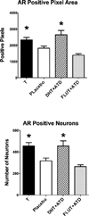
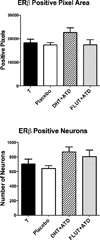
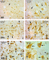
We previously showed that tryptophan hydroxylase 2 (TPH2) and serotonin reuptake transporter (SERT) mRNAs are increased by the androgens, testosterone (T) and dihydrotestosterone (DHT) in serotonin neurons of male macaques. In addition, we observed that serotonin in axons of a terminal region were markedly decreased by aromatase inhibition and lack of estradiol (E) from metabolism of T. These observations implicated androgen receptors (AR) and estrogen receptors (ER) in the transduction of steroid hormone actions in serotonin neurons. Due to the longer treatment period employed, the expression of the cognate nuclear receptors was sought. We used single and double immunohistochemistry to quantitate and phenotypically localize AR, ERα and ERβ in the dorsal raphe of male macaques. Male Japanese macaques (Macaca fuscata) were castrated for 5-7 months and then treated for 3 months with [1] placebo, [2] T, [3] DHT (non-aromatizable androgen) plus ATD (steroidal aromatase inhibitor), or [4] Flutamide (FLUT; androgen antagonist) plus ATD (n = 5/group). After single labeling of each receptor, quantitative image analysis was applied and receptor positive neurons were counted. Double-label of raphe neurons for each receptor plus TPH2 determined whether the receptors were localized in serotonin neurons. There were significantly more AR-positive neurons in T- and DHT+ATD-treated groups (p = 0.0014) compared to placebo or FLUT+ATD-treated groups. There was no difference in the number of positive-neurons stained for ERα or ERβ⋅ Double-immunohistochemistry revealed that serotonin neurons did not contain AR. Rather, AR-positive nuclei were found in neighboring cells that are likely neurons. However, approximately 40% of dorsal raphe serotonin neurons contained ERα or ERβ⋅ In conclusion, the stimulatory effect of androgens on TPH2 and SERT mRNA expression is mediated indirectly by neighboring neurons contain AR. The stimulatory effect of E, derived from T metabolism, on serotonin transport is partially mediated directly via nuclear ERs.
Keywords: Androgen receptors; Estrogen receptors; Macaque; Male; Serotonin.
Copyright © 2015 Elsevier B.V. All rights reserved.
Figures


References
-
- al Saati T, Clamens S, Cohen-Knafo E, Faye JC, Prats H, Coindre JM, Wafflart J, Caveriviere P, Bayard F, Delsol G. Production of monoclonal antibodies to human estrogen-receptor protein (ER) using recombinant ER (RER) Int J Cancer. 1993;55:651–654. - PubMed
-
- Alves SE, Weiland NG, Hayashi S, McEwen BS. Immunocytochemical localization of nuclear estrogen receptors and progestin receptors within the rat dorsal raphe nucleus. J Comp Neurol. 1998;391(3):322–334. - PubMed
-
- Bethea CL. Regulation of progestin receptors in raphe neurons of steroid-treated monkeys. Neuroendocrinology. 1994;60:50–61. - PubMed
-
- Bethea CL, Mirkes SJ, Shively CA, Adams MR. Steroid regulation of tryptophan hydroxylase protein in the dorsal raphe of macaques. Biol Psychiatry. 2000;47:562–576. - PubMed
Publication types
MeSH terms
Substances
Grants and funding
LinkOut - more resources
Full Text Sources
Other Literature Sources
Research Materials

