. 2015 Jul 13;43(12):5880-97.
doi: 10.1093/nar/gkv262. Epub 2015 Apr 23.

Targeting chromatin binding regulation of constitutively active AR variants to overcome prostate cancer resistance to endocrine-based therapies

Affiliations

Targeting chromatin binding regulation of constitutively active AR variants to overcome prostate cancer resistance to endocrine-based therapies

Siu Chiu Chan et al. Nucleic Acids Res. .

Abstract

Androgen receptor (AR) variants (AR-Vs) expressed in prostate cancer (PCa) lack the AR ligand binding domain (LBD) and function as constitutively active transcription factors. AR-V expression in patient tissues or circulating tumor cells is associated with resistance to AR-targeting endocrine therapies and poor outcomes. Here, we investigated the mechanisms governing chromatin binding of AR-Vs with the goal of identifying therapeutic vulnerabilities. By chromatin immunoprecipitation and sequencing (ChIP-seq) and complementary biochemical experiments, we show that AR-Vs display a binding preference for the same canonical high-affinity androgen response elements (AREs) that are preferentially engaged by AR, albeit with lower affinity. Dimerization was an absolute requirement for constitutive AR-V DNA binding and transcriptional activation. Treatment with the bromodomain and extraterminal (BET) inhibitor JQ1 resulted in inhibition of AR-V chromatin binding and impaired AR-V driven PCa cell growth in vitro and in vivo. Importantly, this was associated with a novel JQ1 action of down-regulating AR-V transcript and protein expression. Overall, this study demonstrates that AR-Vs broadly restore AR chromatin binding events that are otherwise suppressed during endocrine therapy, and provides pre-clinical rationale for BET inhibition as a strategy for inhibiting expression and chromatin binding of AR-Vs in PCa.


Figures

Figure 1.
Figure 1.
Outgrowth of AR-V expressing cells in a treatment model of heterogeneous CRPC. (A) Schematic representation of isogenic prostate cancer cell lines R1-AD1 and R1-D567 expressing AR and ARv567es, respectively. (B) Western blots with R1-AD1 and R1-D567 lysates using antibodies specific for the AR NTD and ERK-2 (loading control). (C) Schematic of treatment enrichment experiment. Xenografts were established in intact male mice from a 90%/10% admixture of R1-AD1/R1-D567 cell lines. When tumors reached 100 mm3, mice were castrated and initiated 7-day treatment with 30 mg/kg/day enzalutamide by oral gavage. Biopsies of xenografts were collected at indicated days and the mice were euthanized when tumors reached 1000 mm3. (D) Western blot analysis of protein lysates from tumor tissue collected as in (C) probed with antibodies specific for the AR NTD and ERK-2 (loading control). (E) Multiplex ligation-dependent probe amplification (MLPA) with genomic DNA isolated from pre- and post-implantation samples. Plots illustrate genomic copy number at indicated genomic locations across the AR gene.
Figure 2.
Figure 2.
Genome-wide binding of ARv567es to canonical AREs. (A) Heatmap of ChIP-seq signals ± 3 kb around R1-AD1 AR peak midpoints from three biological replicate experiments for a set of binding sites identified by peak calling with biological replicate 1 data as being common to dihydrotestosterone (DHT)-treated R1-AD1 and vehicle (ethanol, ETH)-treated R1-D567 cells (upper panel), or ‘unique’ to R1-AD1 cells (lower panel). (B) Western blot of chromatin processed for ChIP as in (A) probed with a monoclonal antibody specific for the AR NTD (AR441). (C) Sequence motifs enriched at AR (R1-AD1) and ARv567es (R1-D567) binding sites identified de novo using the Gibbs Motif Sampling approach. (D) Average ChIP-seq tag intensities expressed in mapped reads per base pair per peak normalized per 106 reads from three datasets at binding sites identified as common to R1-AD1 and R1-D567 cells, or ‘unique’ to R1-AD1 cells.
Figure 3.
Figure 3.
Common AR/ARv567es binding sites function as androgen-responsive enhancers. (A) Gene track view of ChIP-seq data at the FASN locus. Common AR/ARv567es binding sites (ARBS) are indicated. Data are from cells treated with dihydrotestosterone (DHT) or vehicle (ethanol, ETH) as indicated. (B) Gene track view of ChIP-seq data at the TSC2 locus. A common AR/ARv567es binding site in TSC2 exon 37 is indicated. (C) Validation of androgen-mediated recruitment of AR to FASN and TSC2 ARBSs in R1-AD1 cells by ChIP-qPCR. (D) Validation of constitutive ARv567es binding to FASN and TSC2 ARBSs in R1-D567 cells by ChIP-qPCR. (E) FASN and TSC2 ARBSs were tested for enhancer response to the synthetic androgen mibolerone (MIB) by luciferase reporter assay in R1-AD1 cells.
Figure 4.
Figure 4.
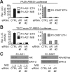
Common AR/ARv567es binding sites are AR/ARv567es-dependent enhancers. (A) AR and ARv567es responsiveness of FASN ARBSI was tested by luciferase reporter assays in R1-AD1 and R1-D567es cells transfected with control (CTRL) siRNA or two separate siRNAs targeting AR. Cells were treated with 1 nM mibolerone (MIB) or ethanol (ETH) as vehicle control as indicated. (B) AR and ARv567es responsiveness of TSC2 exon 37 ARBS was assessed as in (A). (C) Western blots with R1-AD1 and R1-D567 lysates transfected as in (A) and (B) using antibodies specific for the AR NTD and ERK-2 (loading control).
Figure 5.
Figure 5.
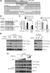
Common AR/ARv567es binding sites regulate endogenous transcriptional outcomes. (A) Schematic of TALENs targeted to an androgen response element (ARE) in FASN ARBSI and examples of mutations recovered by PCR from TALEN-transfected R1-AD1 and R1-D567 cells. (B) T7E1 endonuclease assays evaluating frequency of mutations (assessed by T7E1 cleavage efficiency) in TALEN-transfected R1-AD1 and R1-D567 cells. (C) Quantitative RT-PCR analysis of FASN mRNA levels in R1-AD1 and R1-D567 cells transfected with TALENs targeting FASN AREI or control (CTRL) TALENs targeting an alternate genomic site. Cells were treated with dihydrotestosterone (DHT) or ethanol (ETH) as vehicle control as indicated. (D) Western blots were performed with lysates of R1-AD1 (left) and R1-D567 (right) cells transfected with control (CTRL) siRNA or two separate siRNAs targeting AR and treated with DHT or ETH as vehicle control as indicated. Blots were probed with antibodies specific for the COOH-terminal domain of TSC2, the AR NTD or ERK-2 (loading control). Long and short forms of TSC2 are indicated. (E) Western blots were performed with lysates of LNCaP cells transduced with lentivirus encoding GFP (control) or ARv567es and treated with a range of DHT concentrations (0.1, 1.0, 10 nM) or ethanol (‘–’, vehicle control) as indicated. Blots were probed as in (D).
Figure 6.
Figure 6.
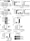
ARv567es binds canonical AREs through a dimerization-dependent mechanism. (A) ARE point mutations introduced in FASN ARBSI-LUC. (B) Activities of constructs illustrated in (A) were tested in R1-AD1 and R1-D567 cells by luciferase assay. Cells were treated with 1 nM mibolerone (MIB) or ethanol (ETH) as vehicle control as indicated. (C) ARE point mutations introduced in TSC2 exon 37-LUC. (D) Activities of constructs illustrated in (C) were evaluated by luciferase assay as in (B). (E) Western blot of lysates from R1-AD1 cells transfected with HA-GFP and HA-AR-V7 for the ChIP experiment shown in (F). (F) Constitutive recruitment of HA-tagged AR-V7 to the FASN ARBS1 site in transfected R1-AD1 cells was tested by ChIP-PCR. Data represent fold enrichment of PCR signal in ChIP DNA isolated using an HA-directed antibody versus non-specific IgG control (which was arbitrarily set to 1). (G) Activities of constructs illustrated in (A) were tested by luciferase assay using LNCaP cells transfected with an ARv567es expression vector and treated with 1 nM mibolerone (MIB) or ethanol (ETH, vehicle) as indicated. (H) Activities of constructs illustrated in (A) were tested by luciferase assay using LNCaP cells transfected with an AR-V7 expression vector exactly as described in (G). (I) Transcriptional activities of wild-type and A596T/S597T D-box mutant versions of ARv567es and AR-V7 were tested in LNCaP cells by luciferase assay as described in (G). (J) DNA duplex pull-down assays were performed by incubating biotinylated FASN AREI DNA duplexes harboring core sequences shown in A with cellular extracts from R1-AD1 and R1-D567 cells.
Figure 7.
Figure 7.
AR-Vs bind a canonical ARE with lower affinity than full-length AR. (A) Representative western blot of lysates from COS-7 cells transfected with plasmids encoding AR, ARv567es and AR-V7 for EMSA experiments. The blot was probed with an antibody specific for the AR NTD. (B) Binding of AR, ARv567es, and AR-V7 to an IRD700-labeled FASN AREI DNA duplex was assessed by electrophoretic mobility shift assay (EMSA). Supershifts were achieved by adding an antibody specific for the AR NTD to binding reactions as indicated. (C) EMSAs were performed as in (B). Labeled FASN AREI duplexes were titrated against a fixed 10 μg of nuclear extract. Apparent equilibrium dissociation constants (Kd) are defined as the concentration of FASN AREI duplex required to achieve half-maximum binding. (D) EMSAs were performed as in (B). Unlabeled competitor DNA duplexes harboring wild-type or mutant FASN ARE1 core sequences shown in Figure 6A were added at 5× and 100× molar excess as indicated.
Figure 8.
Figure 8.
BET inhibitors repress expression of AR and ARv567es in prostate cancer cells. (A) Binding of full-length AR or ARv567es to FASN ARBS1 was tested in R1-AD1 or R1-D567 cells, respectively, treated with vehicle (DMSO) or JQ1 (0.5 mM) in the presence or absence of 1 nM dihydrotestosterone (DHT) as indicated. (B) Western blots with antibodies specific for the AR NTD, AR-V7 or ERK-2 (loading control) with lysates from indicated PCa cell lines treated 24 h with vehicle (DMSO) or JQ1 (doses: 0.1, 0.5, 1, 5, 10 μM) in medium containing 10% CSS. (C) Quantitative RT-PCR analysis of total AR mRNA levels in PCa cell lines treated as in (B). (D) Western blots with antibodies specific for the AR NTD or β-actin (loading control) with lysates from LNCaP cells treated 24 h with vehicle (DMSO) or active S(+) or inactive R(–) JQ1 stereoisomers as indicated. (E) Gene track view of BRD2, BRD3, BRD4 and H3K27Ac ChIP-seq data at the AR locus. Data were obtained from NCBI Gene Expression Omnibus (GEO), representing VCaP cells treated with 0.5 μM JQ1 or vehicle control (DMSO) (GSE27823, (28)) or DHT-treated LNCaP cells (GSE27823, (29)). The FDR-adjusted P value signifying JQ1-mediated loss of BRD2 binding at this site in VCaP cells was derived using diffReps (34). (F) Binding of BRD2 to the AR 5′utr region was tested in R1-AD1 or R1-D567 cells treated with vehicle (DMSO) or JQ1 (0.5 μM) as indicated.
Figure 9.
Figure 9.
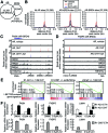
BET inhibitors coordinately suppress transcriptional activation and repression of AR and ARv567es target genes by reducing overall AR chromatin occupancy. (A) Venn diagram representing AR and BRD4 chromatin occupancy in VCaP cells. Occupancy data were obtained from a previous study (28). (B) Average ChIP-seq tag intensities expressed in mapped reads per base pair per peak normalized per 106 reads from three datasets (VCaP cells treated with vehicle, DHT, or DHT and JQ1) obtained from NCBI GEO (GSE27823, (28)). (C) Gene track views of AR, BRD2, BRD3, BRD4 and H3K27Ac ChIP-seq data obtained from NCBI GEO (GSE27823, (28)) at the FASN locus. AR binding sites (ARBS) classified as co-occupied by AR and BRD4 (AR+BRD4) are indicated. (D) Gene track views of the FKBP5 locus developed as in (C). An ARBS classified as occupied by AR but not BRD4 (AR-BRD4) is indicated. (E) Gene set enrichment analysis demonstrating that AR transcriptional activity in R1-AD1 and LNCaP cells, and ARv567es transcriptional activity in R1-D567 cells, is negatively enriched for a set of I-BET762-induced genes. (F) Quantitative RT-PCR analysis of FKBP5, FASN and LIMA1 mRNA expression in R1-AD1 and R1-D567 cells treated with combinations of vehicle (DMSO and ethanol, ETH), 0.5 μM JQ1 or 1 nM DHT as indicated for 24 h.
Figure 10.
Figure 10.
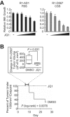
Inhibition of AR-V-driven PCa cell growth by JQ1. (A) Growth assays of R1-AD1 and R1-D567 cells in medium containing 10% FBS (whole serum) or CSS (steroid-depleted serum) with increasing doses of JQ1 (5 nM, 20 nM, 0.1 μM, 0.5 μM and 2 μM). (B) Subcutaneous R1-D567 xenograft tumors were established in intact male mice. When tumors reached 100 mm3, mice were randomized to treatment (JQ1, n = 7) or control (DMSO, n = 7) groups for 24 days. Top: change in tumor volume from day 7 to day 10 for DMSO versus JQ1 groups. Boxes represent first to third quartiles with median; whiskers represent range. P values were derived using a two-tailed t-test. Bottom: Kaplan–Meier analysis of DMSO versus JQ1 groups, representing time for tumors to reach 1000 mm3. P values were derived using a log-rank (Mantel–Cox) test.

References

    1. Matusik R.J., Jin R.J., Sun Q., Wang Y., Yu X., Gupta A., Nandana S., Case T.C., Paul M., Mirosevich J., et al. Prostate epithelial cell fate. Differentiation. 2008;76:682–698. - PubMed
    1. Kumar-Sinha C., Tomlins S.A., Chinnaiyan A.M. Recurrent gene fusions in prostate cancer. Nat. Rev. Cancer. 2008;8:497–511. - PMC - PubMed
    1. Knudsen K.E., Scher H.I. Starving the addiction: new opportunities for durable suppression of AR signaling in prostate cancer. Clin. Cancer Res. 2009;15:4792–4798. - PMC - PubMed
    1. Logothetis C.J., Gallick G.E., Maity S.N., Kim J., Aparicio A., Efstathiou E., Lin S.H. Molecular classification of prostate cancer progression: foundation for marker-driven treatment of prostate cancer. Cancer Discov. 2013;3:849–861. - PMC - PubMed
    1. Ryan C.J., Tindall D.J. Androgen receptor rediscovered: the new biology and targeting the androgen receptor therapeutically. J. Clin. Oncol. 2011;29:3651–3658. - PubMed

Publication types

MeSH terms

Associated data