. 2015 Apr 24;348(6233):457-60.
doi: 10.1126/science.1261909. Epub 2015 Apr 23.

Ribosome. Mechanical force releases nascent chain-mediated ribosome arrest in vitro and in vivo

Affiliations

Ribosome. Mechanical force releases nascent chain-mediated ribosome arrest in vitro and in vivo

Daniel H Goldman et al. Science. .

Abstract

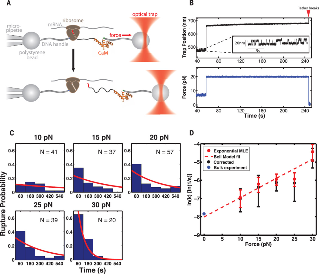
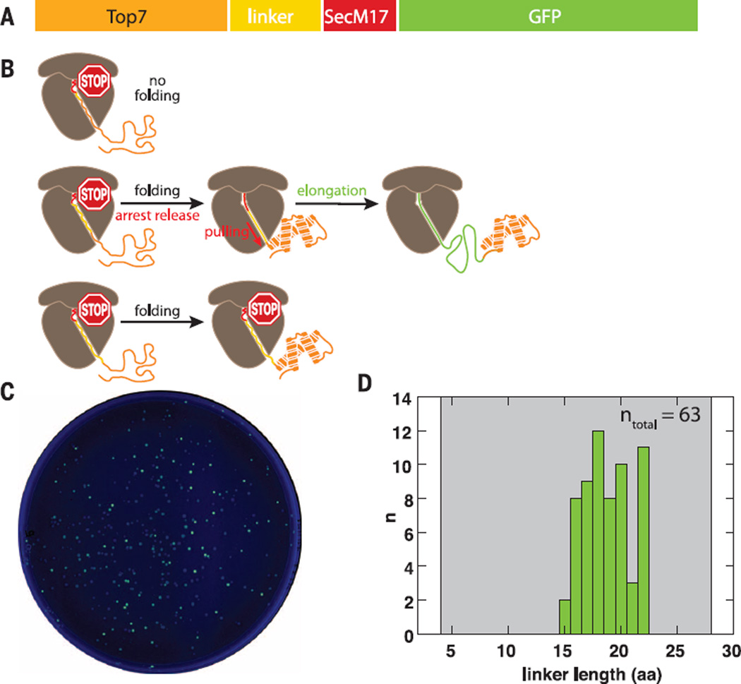
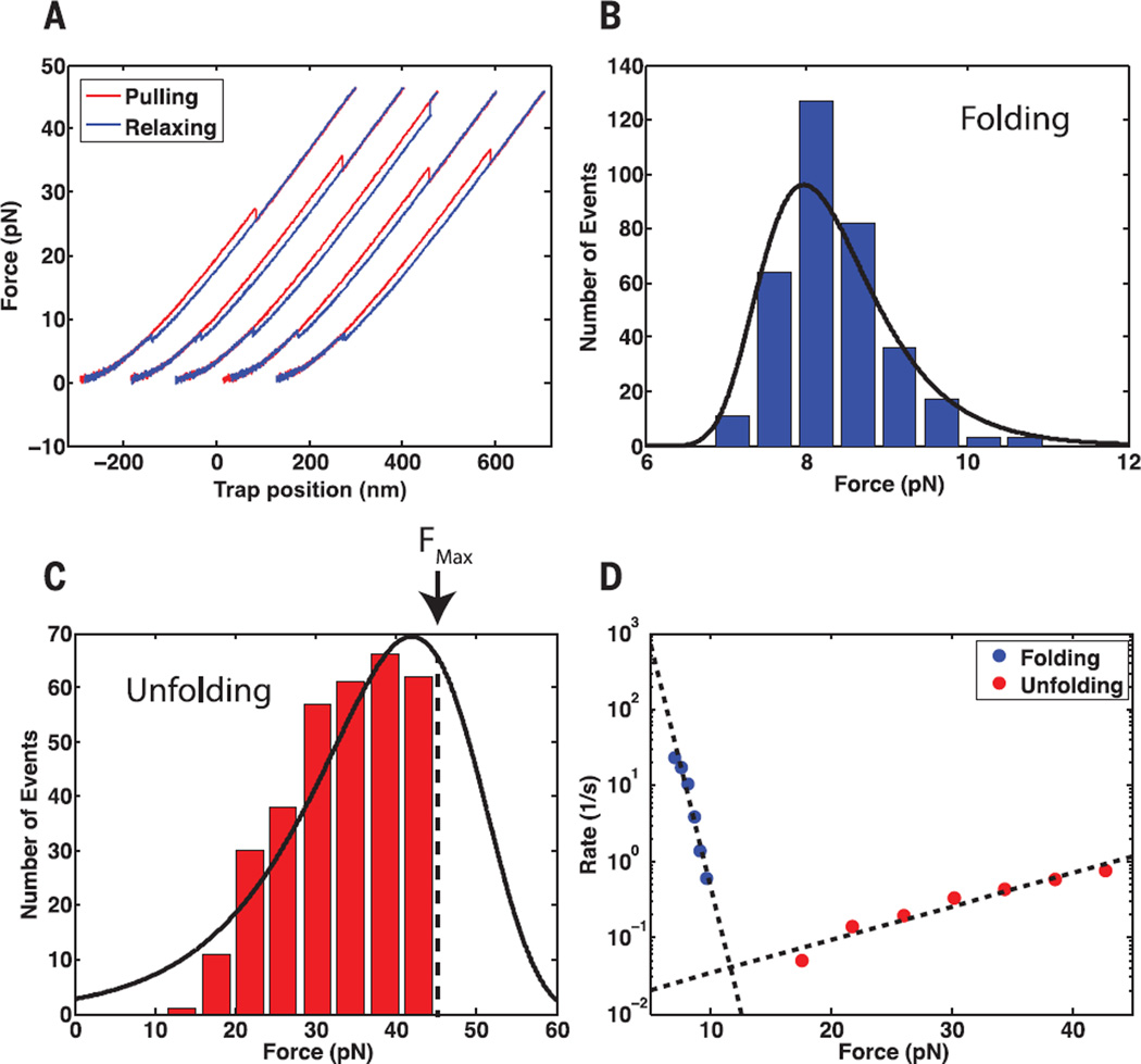
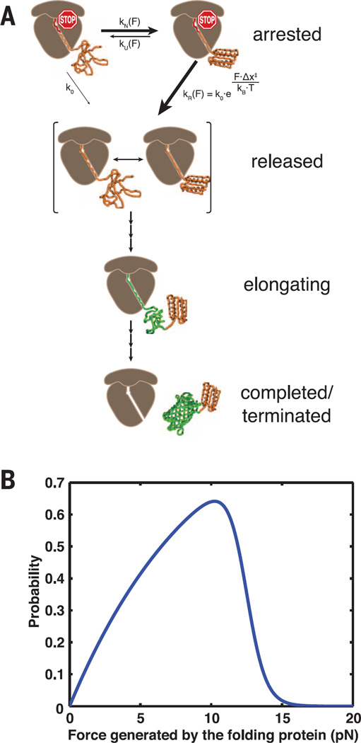
Protein synthesis rates can affect gene expression and the folding and activity of the translation product. Interactions between the nascent polypeptide and the ribosome exit tunnel represent one mode of regulating synthesis rates. The SecM protein arrests its own translation, and release of arrest at the translocon has been proposed to occur by mechanical force. Using optical tweezers, we demonstrate that arrest of SecM-stalled ribosomes can indeed be rescued by force alone and that the force needed to release stalling can be generated in vivo by a nascent chain folding near the ribosome tunnel exit. We formulate a kinetic model describing how a protein can regulate its own synthesis by the force generated during folding, tuning ribosome activity to structure acquisition by a nascent polypeptide.


Figures

Fig. 1
Fig. 1. A direct applied force catalyzes release of SecM-mediated arrest
(A) Experimental setup for optical tweezers experiments. When the nascent chain is transferred to puromycin, the assembly breaks. The structure of CaM was obtained from Protein Data Bank (PDB) ID 1CLL. (B) Example trace for restart experiment. After the “hopping” signature of CaM is observed (inset) at 7 pN, the force is raised to 20 pN. Red arrow: The tether breaks after ~3 min at 20 pN. (C) Restart lifetimes at each force. Red lines: Distributions returned by the right-censoring MLE. (D) Force-dependent rates for restart of SecM-stalled RNCs in the optical tweezers. Rates are determined as shown in (C), with error bars representing 95% CIs returned by the MLE. Red dotted line: Fit of Bell’s model to optical tweezers data. Δx: 0.4 nm (95% CI: 0.1 nm, 0.8 nm) and k0: 3.3 × 10⁻⁴ s−1 (95% CI: 0.5 × 10⁻⁴ s−1, 20 × 10−4 s−1). Black points: Rates determined with a method to account for nonspecific tether rupture (fig. S6). Error determined by bootstrapping. Blue dot: Lifetime obtained from bulk experiment (fig. S3).
Fig. 2
Fig. 2. Nascent protein folding near the ribosome tunnel exit can rescue SecM-mediated stalling
(A) Primary sequence of the construct used in the GFP reporter assay. (B) Schematic illustrating the translation outcome for a short (top), intermediate (middle), and long (bottom) linker. (C) Ultraviolet-illuminated image of colonies transformed with the linker library and grown under inducing conditions. (D) Histogramof linker lengths recovered by sequencing of fluorescent colonies. Gray shaded area: library range.
Fig. 3
Fig. 3. Top7 refolds against an applied mechanical load
(A) Example force ramp cycles for a single Top7 molecule. Pulling is shown in red, relaxing in blue. Successive cycles are offset along the x axis for display purposes. (B and C) Folding and unfolding force distributions, respectively, for Top7 at a pulling speed of 100 nm/s. Black line: Distributions reconstructed from the force-dependent rates in (D). The unfolding-force distribution in (C) is right-censored because the maximum force in pulling experiments was set at 45 pN to avoid tether rupture. (D) Force-dependent rates of folding and unfolding extracted from the distributions in (B and C). Dashed lines: fit of Bell’s model to the force-dependent rates. For folding, Δx: 6 nm (95% CI: 4 nm, 8 nm) and k0: 1 × 10⁶ s−1 (95% CI: 0.04 × 10⁶ s−1, 30 × 106 s−1). For unfolding, Δx: 0.4 nm (95% CI: 0.3 nm, 0.6 nm) and k0: 0.01 s−1 (95% CI: 0.003 s−1, 0.03 s−1) (supplementary materials).
Fig. 4
Fig. 4. Kinetic model for folding-induced release of stalled ribosomes
(A) Kinetic scheme illustrating the pathway to release of translation arrest. The nascent polypeptide can transit reversibly between the native and unfolded states, with rate constants kN(F) and kU(F). Once folded, the nascent protein both generates and experiences a force, “F,” which can drive it either irreversibly to the “released” state, with rate constant kR(F), or back to the unfolded state. In addition, the stall can be released via the irreversible spontaneous process from the unfolded state, with rate constant k0, which is independent of force. PDB: 1QYS and 1EMA. (B) The probability of force-catalyzed stall release is plotted as a function of the folding force.

Comment in

References

    1. Nissley DA, O’Brien EP. JACS. 2014;136:17892–17898. - PubMed
    1. Ito K, Chiba S. Annu. Rev. Biochem. 2013;82:171–202. - PubMed
    1. Deutsch . Regulatory Nascent Polypeptides. Tokyo: Springer; 2014. pp. 61–86.
    1. Wilson DN, Beckmann R. Curr. Opin. Struct. Biol. 2011;21:274–282. - PubMed
    1. Tsai A, Kornberg G, Johansson M, Chen J, Puglisi JD. Cell Rep. 2014;7:1521–1533. - PMC - PubMed

Publication types

MeSH terms