

Controlled induction of human pancreatic progenitors produces functional beta-like cells in vitro
- PMID: 25908839
- PMCID: PMC4516429
- DOI: 10.15252/embj.201591058
Controlled induction of human pancreatic progenitors produces functional beta-like cells in vitro
Abstract
Directed differentiation of human pluripotent stem cells into functional insulin-producing beta-like cells holds great promise for cell replacement therapy for patients suffering from diabetes. This approach also offers the unique opportunity to study otherwise inaccessible aspects of human beta cell development and function in vitro. Here, we show that current pancreatic progenitor differentiation protocols promote precocious endocrine commitment, ultimately resulting in the generation of non-functional polyhormonal cells. Omission of commonly used BMP inhibitors during pancreatic specification prevents precocious endocrine formation while treatment with retinoic acid followed by combined EGF/KGF efficiently generates both PDX1(+) and subsequent PDX1(+)/NKX6.1(+) pancreatic progenitor populations, respectively. Precise temporal activation of endocrine differentiation in PDX1(+)/NKX6.1(+) progenitors produces glucose-responsive beta-like cells in vitro that exhibit key features of bona fide human beta cells, remain functional after short-term transplantation, and reduce blood glucose levels in diabetic mice. Thus, our simplified and scalable system accurately recapitulates key steps of human pancreas development and provides a fast and reproducible supply of functional human beta-like cells.
Keywords: beta‐like cells; diabetes; human embryonic stem cells; insulin‐producing cells, pancreas.
© 2015 The Authors.
Figures
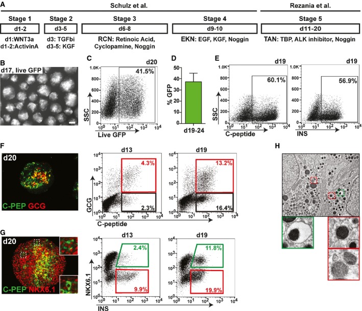
Schematic outlining the differentiation protocol employed. R, retinoic acid; C, cyclopamine; N, Noggin, E, epidermal growth factor; K, keratinocyte growth factor; T, TBP; A, ALK inhibitor.
Micrograph of MEL1INS-GFP cell clusters after 17 days of differentiation demonstrating strong GFP expression (GFP expression in white). Scale bar, 200 μm.
Flow cytometric analysis at day 20 of differentiation showing 41.5% of all cells expressing GFP under the control of the endogenous insulin promoter.
Quantification by flow cytometry of the average percentage of GFP+ cells within differentiated cultures after 19–24 days. n = 7. Values are average ± standard deviation (SD).
Flow cytometric analysis of intracellular human-specific C-peptide (C-PEP) and insulin (INS) shows comparable percentages of C-PEP− and INS+ cells.
Immunofluorescence staining for C-PEP and glucagon (GCG), and flow cytometric quantification of GCG+/C-PEP+ (red gate) and GCG−/C-PEP+ (black gate) populations at days 13 and 19 of differentiation.
Immunofluorescence staining for C-PEP and NKX6.1, and flow cytometric quantification of NKX6.1+/INS+ (green gate) and NKX6.1−/INS+ (red gate) populations at day 13 and 19. Immunofluorescence insets show two distinct phenotypes for C-PEP+ cells (NKX6.1+ and NKX6.1−). A robust INS/NKX6.1 double-positive population is only detected at day 19.
Transmission electron microscopy of day 20 clusters. Cells contain both secretory vesicles with electron-dense cores surrounded by electron-light halos (green box), akin to bona fide beta cell vesicles, as well as other granules similar to those found in non-beta pancreatic cells (red boxes).
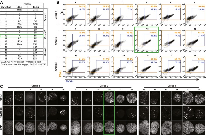
A–C Pancreatic progenitor marker expression at day 9.5 after treatment with conventional differentiation factors alone or in different combinations. Treatments consisted of combinations of cyclopamine (C), Noggin (N), and retinoic acid (R) during days 6–8 followed by subdivision of each condition into three treatment groups during day 9–9.5. Group 1: continuation of day 6–8 treatment; Group 2: treatment with EGF and KGF (EK); Group 3: treatment with EGF, KGF, and Noggin (EKN). The condition selected for further studies (10) is marked with a green box. Data shown are representatives of results obtained in two independent experiments. (A) Table detailing 18 different culture conditions that were evaluated. (B) Quantification of PDX1 (orange gate) and NKX6.1 (blue gate) protein-expressing cells in individual conditions after 9.5 days of differentiation. (C) NKX6.1 and NEUROG3 protein expression assessed by whole-mount staining of differentiated clusters at 9.5 days. Note robust NEUROG3 expression in all clusters exposed to N (conditions 3, 6, 9, and 12–18).
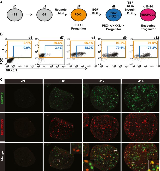
Schematic outlining a simplified differentiation strategy for the controlled, stepwise generation of pancreatic progenitor cell types.
Time-course flow cytometric analysis illustrates the efficient generation of PDX1+ progenitor (orange gate) and PDX1+/NKX6.1+ progenitor (blue gate) populations. Data from one of three independent experiments with similar results are shown.
Immunofluorescence analysis of sections from differentiated clusters at indicated time points stained for human NKX2.2 (green) and NEUROG3 (red). Insets show NEUROG3/NKX2.2 double-positive cells. Data from one of three independent experiments with similar results are shown.
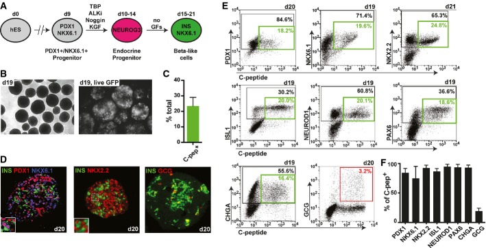
Schematic outlining a simplified differentiation strategy for the controlled, stepwise generation of pancreatic progenitor and subsequent endocrine cell types. GFs, growth factors.
Micrographs of differentiated clusters at day 19 under light microscopy (left picture) or fluorescent microscopy showing prominent GFP expression (right picture; GFP expression shown in white).
Quantification of the percentage of human C-peptide-positive cells at day 19–21. Values are average ± SD. n = 7 independent experiments.
Immunofluorescence stainings of differentiated clusters at day 20 for insulin (INS), PDX1, NKX6.1, NKX2.2, and glucagon (GCG). One of four experiments with similar outcome is shown.
Representative flow cytometry plots depicting co-expression of pancreatic markers PDX1, NKX6.1, NKX2.2, ISL1, NEUROD1, PAX6, chromogranin A (CHGA), and GCG with human C-peptide at indicated time points. Black gates mark percentage of total cells positive for indicated marker on y-axis. Green gates mark percentage of double-positive beta-like cells. The red gate marks percentage of INS+/GCG+ bihormonal cells.
Flow cytometric quantification of C-peptide-positive beta-like cells co-expressing markers in (E). A high percentage of beta-like cells co-express all genes usually found in beta cells, but not the hormone GCG. Values are average ± SD. n = 4 for PDX1, n = 19 for NKX6.1, n = 4 for NKX2.2, n = 9 for ISL1, n = 9 for NEUROD1, n = 5 for PAX6, n = 6 for CHGA, and n = 5 for GCG. Analysis was performed at days 15–21 of differentiation.
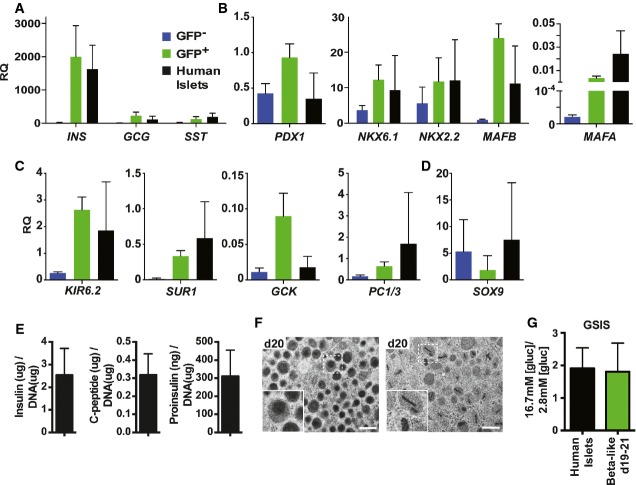
A–D Quantitative PCR analysis of selected gene transcripts in sorted GFP+ beta-like cells (green bars), GFP− populations (blue bars) and human islet preparations (black bars). Results shown relative to the endogenous control GAPDH. RQ, relative quantification. Values are average ± SD. n = 4 independent experiments for hESC-derived cell populations at days 19–20 and n = 3 for human islets.
E Insulin, human C-peptide, and proinsulin content relative to DNA in beta-like cells at day 19. Data presented are average ± standard error (n = three independent experiments, technical duplicates).
F Transmission electron microscopy images of beta-like cells at day 20. One of three experiments with similar results is shown. Scale bar, 500 nm. Insets represent secretory vesicles akin to granules present in bona fide human beta cells.
G Glucose-stimulated insulin secretion (GSIS) of human islets and beta-like cells at days 19–20. Y-axis indicates ratio of insulin secreted in low glucose conditions to that secreted in high glucose conditions. Values are average ± standard deviation (SD). n = 3 for human islets and n = 10 for beta-like cells.
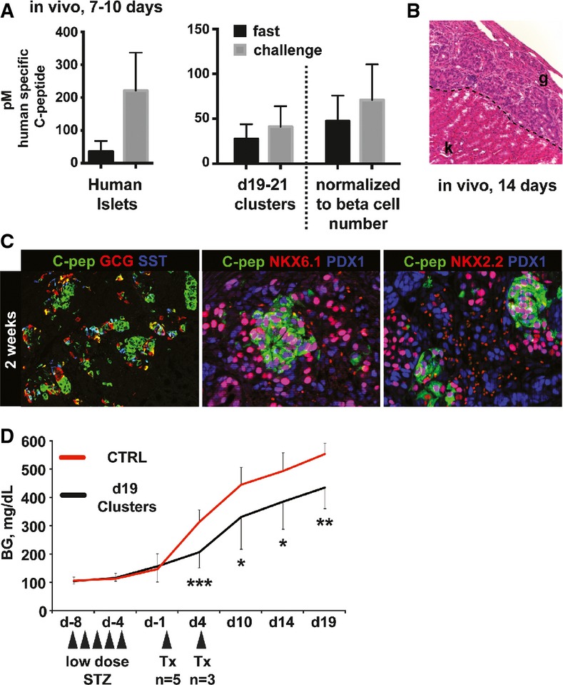
Levels of circulating human C-peptide measured in sera of mice 7–10 days after transplantation with either 4,000 human islets or 5.0 × 10⁶ direct differentiated cells (containing approximately 1.15 × 10⁶ beta-like cells). Fasting and challenge sera were collected following an overnight fast and 1 h after intraperitoneal glucose challenge, respectively. Dashed line separates raw data from serum C-peptide measurements normalized to the number of beta cells present in each human islet graft (4,000 human islets transplanted each containing ˜1,000 cells, approximately 50% of which are beta cells, hence 2.0 × 10⁶ beta cells present in grafts total). n = 5 for human islets and n = 12 for hESC-derived grafts.
Hematoxylin and eosin staining of day 14 graft. k, kidney; g, graft. Representative data from one of three mice are shown.
Immunofluorescence staining of differentiated hESC grafts 2 weeks post-transplantation for human C-peptide, glucagon (GCG), somatostatin (SST), PDX1, NKX6.1, and NKX2.2. Representative data from one of three mice are shown.
Blood glucose (BG) levels of mice treated with streptozotocin to ablate endogenous beta cells (STZ) followed by transplantation (Tx) of beta-like cell containing clusters either at day 0 or at day 4 as indicated (n = 8, two independent differentiation experiments). Values are average ± SD. Statistical significance was calculated using two-tailed t-test. *P < 0.05, **P < 0.01, and ***P < 0.001. Control (CTRL) group: 6–9 animals.
Comment in
-
Simply the right time to turn on insulin.EMBO J. 2015 Jul 2;34(13):1740-2. doi: 10.15252/embj.201591894. Epub 2015 May 13. EMBO J. 2015. PMID: 25971777 Free PMC article.
References
-
- Barton FB, Rickels MR, Alejandro R, Hering BJ, Wease S, Naziruddin B, Oberholzer J, Odorico JS, Garfinkel MR, Levy M, Pattou F, Berney T, Secchi A, Messinger S, Senior PA, Maffi P, Posselt A, Stock PG, Kaufman DB, Luo X, et al. Improvement in outcomes of clinical islet transplantation: 1999–2010. Diabetes Care. 2012;35:1436–1445. - PMC - PubMed
-
- Bouwens L, Houbracken I, Mfopou JK. The use of stem cells for pancreatic regeneration in diabetes mellitus. Nat Rev Endocrinol. 2013;9:598–606. - PubMed
-
- Chen S, Borowiak M, Fox JL, Maehr R, Osafune K, Davidow L, Lam K, Peng LF, Schreiber SL, Rubin LL, Melton D. A small molecule that directs differentiation of human ESCs into the pancreatic lineage. Nat Chem Biol. 2009;5:258–265. - PubMed
-
- D'Amour KA, Agulnick AD, Eliazer S, Kelly OG, Kroon E, Baetge EE. Efficient differentiation of human embryonic stem cells to definitive endoderm. Nat Biotechnol. 2005;23:1534–1541. - PubMed
