. 2015 Jun 1;128(11):2191-201.
doi: 10.1242/jcs.170316. Epub 2015 Apr 23.

Dendrite arborization requires the dynein cofactor NudE

Affiliations

Dendrite arborization requires the dynein cofactor NudE

Ashley L Arthur et al. J Cell Sci. .

Abstract

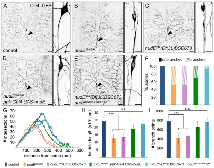
The microtubule-based molecular motor dynein is essential for proper neuronal morphogenesis. Dynein activity is regulated by cofactors, and the role(s) of these cofactors in shaping neuronal structure are still being elucidated. Using Drosophila melanogaster, we reveal that the loss of the dynein cofactor NudE results in abnormal dendrite arborization. Our data show that NudE associates with Golgi outposts, which mediate dendrite branching, suggesting that NudE normally influences dendrite patterning by regulating Golgi outpost transport. Neurons lacking NudE also have increased microtubule dynamics, reflecting a change in microtubule stability that is likely to also contribute to abnormal dendrite growth and branching. These defects in dendritogenesis are rescued by elevating levels of Lis1, another dynein cofactor that interacts with NudE as part of a tripartite complex. Our data further show that the NudE C-terminus is dispensable for dendrite morphogenesis and is likely to modulate NudE activity. We propose that a key function of NudE is to enhance an interaction between Lis1 and dynein that is crucial for motor activity and dendrite architecture.

Keywords: Dendrite patterning; Drosophila; Dynein; Microtubules; Nde1; Ndel1; NudE.


Conflict of interest statement

Competing interests

The authors declare no competing or financial interests.

Figures

Fig. 1.
Fig. 1.
Loss of NudE disrupts dendrite and axon morphogenesis. (A–E) Representative images of class IV ddaC neurons illuminated by expression of ppk-CD4–GFP in live intact 3rd instar larvae. Arrowhead indicates axon. Scale bars: 50 µm (left-hand images); 10 µm (right-hand images showing magnified views of axon). (F–I) Quantification of axon branching (F), dendrite patterning (G), dendrite length (H) and dendrite branching (I) phenotypes. For nudE39A/39A ppk-Gal4 UAS-nudE neurons, dendrite length was significantly different from that of control and nudE39A/39A neurons (P=0.003 and P=0.0004, respectively); the number of branch points was also significantly different from those of control and nudE39A/39A neurons (P=0.05 and P=0.002, respectively). Error bars indicate s.d., ***P=0.001–0.0001; n.s., not significant; n=8 neurons for all genotypes except for nudE39A/39A n=9. The color code for the genotypes is shown at the bottom of the figure.
Fig. 2.
Fig. 2.
Loss of NudE enhances the dynein loss-of-function dendrite morphogenesis phenotype. (A–D) Representative images of class IV ddaC neurons illuminated by expression of ppk-CD4–GFP in live intact 3rd instar larvae. Dendrite patterning was mildly disrupted in ppk-Gal4 UAS-dlic-RNAi larvae (A). The expression of dcr enhances the dendrite arborization phenotype caused by reducing levels of Dlic (C). Reducing levels of NudE and Dlic at the same time reduces dendrite growth and branching, similar to ppk-Gal4 UAS-dlic-RNAi UAS-dcr (D). Arrowheads indicate axons. Scale bars: 50 µm.
Fig. 3.
Fig. 3.
NudE colocalizes with Golgi outposts in dendrites and prevents them from entering axons. (A,A′) Representative image of a ddaC neuron co-expressing CD4–GFP (A) and Cherry–NudE (A′). Arrowhead indicates axon. Inset in A′ shows a magnified view of an axon, arrows indicate several Cherry–NudE particles. (B) Representative kymograph showing the motility of Cherry–NudE (B, red channel in B″) and the Golgi marker ManII–GFP (B′, green channel in B″) in dendrites. Cell body is to the left. (C) Quantification of Cherry–NudE and ManII–GFP movement and colocalization in dendrites (the distances that Cherry–NudE and ManII–GFP particles travel in the anterograde or retrograde direction are similar, data not shown). (D,E) Representative images (D) and quantification (E) of Golgi outposts in the axons of control (ppk-Gal4 UAS-ManII–GFP, left panel in D) and nudE39A/39A (right panel in D) neurons. Arrowheads indicate Golgi outposts. Error bars indicate s.d., **P=0.01–0.001, n=19 control axons, n=15 nudE39A/39A axons. Scale bars: 50 µm (A); 10 µm (D, inset of A′); 5 µm (x axis of B″); 30 s (y axis of B″).
Fig. 4.
Fig. 4.
Loss of NudE affects microtubule growth and the polarity of axonal but not dendritic microtubules. (A) Kymographs generated from representative movies of EB1–GFP comets present in the axons (top) and dendrites (bottom) of neurons in live intact 3rd instar larvae. ppk-EB1–GFP control (left) and nudE39A/39A (right) neurons are shown. The cell body is to the left in all kymographs. Scale bars: 5 µm (x axis); 30 s (y axis). (B) Quantification of EB1–GFP comet frequency in axons (top) and dendrites (bottom). Comet frequency reflects the number of growing microtubules (control axons, n=13 neurons, which includes a total of 58 comets; nudE39A/39A axons, n=11 neurons, which includes a total of 156 comets; control dendrites, n=24 dendrite segments in nine neurons, which includes a total of 247 comets; nudE39A/39A dendrites, n=32 dendrite segments in eight neurons, which includes a total of 540 comets). Boxes represent first and third quartiles (median indicated by line) and whiskers indicate minimum and maximum values. (C) Quantification of the direction that EB1–GFP comets traveled, which reflects the polarity of microtubules in axons (top) and dendrites (bottom) (control axons, n=11 axons with 135 comets; control dendrites, n=12 dendrites with 323 comets; nudE39A/39A axons, n=9 axons with 115 comets; nudE39A/39A dendrites, n=8 dendrites with 540 comets). Error bars indicate s.d., **P=0.01–0.001, ***P=0.001–0.0001, ****P<0.0001; n.s., not significant.
Fig. 5.
Fig. 5.
γ-tubulin-mediated microtubule nucleation is not responsible for the change in axonal microtubule polarity that is caused by the loss of NudE. (A,B) The polarity of axonal microtubules was determined using EB1–GFP, the comet trajectories of which are plotted in kymographs. The cell body is to the left in all kymographs. Scale bars: 5 µm (x axis); 30 s (y axis). (C) Quantification of the direction that EB1–GFP comets traveled in axons. γTub23CA14-9/A15-2, n=16 axons with 156 comets. γTub23CA15-2/A14-9; nudE39A/39A, n=12 axons with 193 comets. The percentage of comets traveling anterograde and retrograde in the axons of nudE39A/39A and γTub23CA15-2/A14-9; nudE39A/39A neurons was significantly different from control (****P<0.0001 for both genotypes). n.s., not significant.
Fig. 6.
Fig. 6.
The NudE N-terminus is sufficient for normal dendrite arborization. (A,B) Representative images of class IV ddaC neurons illuminated through expression of ppk-CD4–GFP in live intact 3rd instar larvae. Arrowhead indicates axons. Scale bars: 50 µm. (C,D) Dendrite arborization was quantified using Sholl analysis (C) and by measuring dendrite length (D). Error bars indicate s.d., **P=0.01–0.001, ***P=0.001–0.0001; n.s., not significant; n=8 neurons for all genotypes, except n=9 for nudE39A/39A.
Fig. 7.
Fig. 7.
Overexpressing Lis1 rescues abnormal dendrite arborization that is caused by the loss of NudE. (A–D) Representative images of ddaC neurons illuminated through expression of ppk-CD4–GFP in live intact 3rd instar larvae. Arrowheads indicate axons. Scale bars: 50 µm. (E,F) Dendrite arborization was quantified using Sholl analysis (E) and by measuring dendrite length (F). Error bars indicate s.d., ***P=0.001–0.0001, n.s., not significant; n=8 neurons for all genotypes, except n=9 for nudE39A/39A.

References

    1. Andersen R., Li Y., Resseguie M. and Brenman J. E. (2005). Calcium/calmodulin-dependent protein kinase II alters structural plasticity and cytoskeletal dynamics in Drosophila. J. Neurosci. 25, 8878-8888. 10.1523/JNEUROSCI.2005-05.2005 - DOI - PMC - PubMed
    1. Baas P. W. and Mozgova O. I. (2012). A novel role for retrograde transport of microtubules in the axon. Cytoskeleton (Hoboken) 69, 416-425. 10.1002/cm.21013 - DOI - PubMed
    1. Bradshaw N. J., Hennah W. and Soares D. C. (2013). NDE1 and NDEL1: twin neurodevelopmental proteins with similar ‘nature’ but different ‘nurture’. Biomol Concepts 4, 447-464. 10.1515/bmc-2013-0023 - DOI - PMC - PubMed
    1. Chabin-Brion K., Marceiller J., Perez F., Settegrana C., Drechou A., Durand G. and Poüs C. (2001). The Golgi complex is a microtubule-organizing organelle. Mol. Biol. Cell 12, 2047-2060. 10.1091/mbc.12.7.2047 - DOI - PMC - PubMed
    1. Chansard M., Hong J. H., Park Y. U., Park S. K. and Nguyen M. D. (2011). Ndel1, Nudel (Noodle): flexible in the cell? Cytoskeleton (Hoboken) 68, 540-554. 10.1002/cm.20532 - DOI - PubMed

Publication types

MeSH terms

LinkOut - more resources