. 2015 Jun 1;128(11):2096-105.
doi: 10.1242/jcs.166009. Epub 2015 Apr 23.

Induction of stable ER-plasma-membrane junctions by Kv2.1 potassium channels

Affiliations

Induction of stable ER-plasma-membrane junctions by Kv2.1 potassium channels

Philip D Fox et al. J Cell Sci. .

Abstract

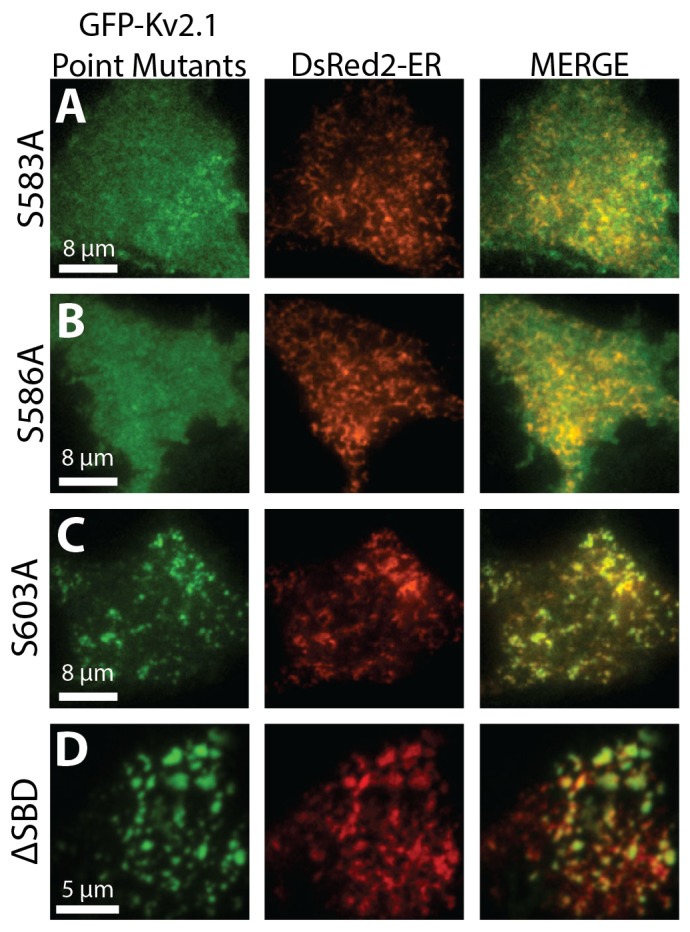
Junctions between cortical endoplasmic reticulum (cER) and the plasma membrane are a subtle but ubiquitous feature in mammalian cells; however, very little is known about the functions and molecular interactions that are associated with neuronal ER-plasma-membrane junctions. Here, we report that Kv2.1 (also known as KCNB1), the primary delayed-rectifier K(+) channel in the mammalian brain, induces the formation of ER-plasma-membrane junctions. Kv2.1 localizes to dense, cell-surface clusters that contain non-conducting channels, indicating that they have a function that is unrelated to membrane-potential regulation. Accordingly, Kv2.1 clusters function as membrane-trafficking hubs, providing platforms for delivery and retrieval of multiple membrane proteins. Using both total internal reflection fluorescence and electron microscopy we demonstrate that the clustered Kv2.1 plays a direct structural role in the induction of stable ER-plasma-membrane junctions in both transfected HEK 293 cells and cultured hippocampal neurons. Glutamate exposure results in a loss of Kv2.1 clusters in neurons and subsequent retraction of the cER from the plasma membrane. We propose Kv2.1-induced ER-plasma-membrane junctions represent a new macromolecular plasma-membrane complex that is sensitive to excitotoxic insult and functions as a scaffolding site for both membrane trafficking and Ca(2+) signaling.

Keywords: Ion channels; Membrane contact sites; Sub-surface cisterns.


Conflict of interest statement

Competing interests

The authors declare no competing or financial interests.

Figures

Fig. 1.
Fig. 1.
Clustering of Kv2.1 remodels the cER. Two HEK cells, outlined in white, that had been transfected with the luminal ER marker DsRed2-ER (A) and GFP–Kv2.1 (B) were imaged using TIRF microscopy. The bottom cell has large GFP–Kv2.1 clusters, whereas the top left cell has no Kv2.1 expression. Both cells exhibit similar intensities of DsRed2-ER. (C) Enlargement of the region in the yellow box from A. Note the mesh-like pattern of the cER, which comprises tubules and bright puncta (yellow arrow). (D) Enlargement of the green box in A. Note the absence of mesh-like tubules in favor of large planar cER structures (white arrow). (E) Histogram of the pixel intensities of the entire cER (red boxes) and the Kv2.1-cluster-associated cER (black boxes) obtained from imaging DsRed2-ER in TIRF and averaged over six cells (mean±s.e.m.). The brightest cER pixels were almost exclusively present underneath Kv2.1 clusters, suggesting close proximity to the plasma membrane. (F) Representative 7-DIV rat hippocampal neuron that had been transfected with an ER marker comprising the membrane domain of cytochrome B5 (CYB5A) fused to GFP (GFP–CB5) and imaged in TIRF. The majority of neurons imaged at this time in culture had apparent tubular cER and small punctate cER but lacked larger cER structures. (G,H) A representative 7-DIV rat hippocampal neuron that had been transfected with GFP–CB5 (G) and Kv2.1-LoopBAD, which comprises an extracellular biotin tag (H, surface labeled with streptavidin–Alexa594). The large, Kv2.1-associated cER structures (G, GFP–CB5) were strikingly different than anything present in 7-DIV neurons that lacked ectopic expression of Kv2.1.
Fig. 2.
Fig. 2.
Kv2.1 clustering induces the formation of true ER–plasma-membrane junctions. Immuno-gold labeling and electron microscopy were used to visualize the ultrastructural features of Kv2.1-remodeled cER. (A) Electron microscopy micrographs of cER from mock-transfected HEK cells. The four micrographs were obtained from cells under identical conditions. Top left panel shows a long stretch of cER that fails to make the consistent close contact that is a hallmark of ER–plasma-membrane junctions. Black arrows in the top-right and bottom panels point to endogenous ER–plasma-membrane junctions, which tended to be ∼200 nm in length. (B) Two representative electron microscopy micrographs of long ER–plasma-membrane junctions (∼1 μm) in a Kv2.1–HA transfected HEK cell. Kv2.1–HA channels on the cell surface labeled with 10-nm- and 20-nm gold. Scale bars: 200 nm (all electron microscopy micrographs). (C) Bar graphs summarizing the effect of Kv2.1 expression on ER–plasma-membrane junctions in HEK cells. The left-hand graph illustrates the average length of ER–plasma-membrane junctions in cells positive for Kv2.1 by using immuno-gold labeling (n=139 junctions averaged from 20 cells) versus mock-transfected cells (n=132 junctions averaged from 36 cells). The right-hand graph shows the percentage of the plasma membrane that was associated ER junctions in cells that were positive for immuno-gold labeling of Kv2.1 (n=20 cells) versus mock-transfected cells (n=36 cells), mean±s.e.m., *P<0.01.
Fig. 3.
Fig. 3.
Clustering of Kv2.1 and formation of ER–plasma-membrane junctions. Transfected HEK cells expressing DsRed2-ER (A) and GFP–Kv2.1 (B) were removed from the incubator, the growth medium was replaced with imaging saline, and the cells were placed on the TIRF microscope stage. Frequently, cells expressing GFP–Kv2.1 initially lacked clusters (A, left panel) and the cER was largely tubular in appearance (B, left panel). Over the next 20 min of imaging, Kv2.1 clusters formed (A, right panel) and the cER remodeled from tubules into larger, planar structures (B, right panel). The time from the start of imaging is shown in the top right of the images in A. (C) The average normalized intensities from six GFP–Kv2.1 clusters (green line) and six cluster-associated cER puncta (red line) during Kv2.1 re-clustering (mean±s.e.m.). For intensity measurements, the locations of Kv2.1 clusters and associated cER puncta were automatically tracked using a centroid-based algorithm implemented in LabView. In this cell, the fluorescence intensities of Kv2.1 clusters and cER increased in concert with one another (see supplementary material Movie 1).
Fig. 4.
Fig. 4.
Kv2.1 clustering enhances the stability of underlying cER. (A) Two HEK cells co-transfected with DsRed2-ER (A, single channel) and GFP–Kv2.1 (B, single channel), imaged in TIRF. The cell on the bottom left has abundant Kv2.1 clusters, whereas the cell in the top right is completely devoid of clustered Kv2.1. ‘(D)’ and ‘(E)’ refer to the respective figure panels. (C) A merge of the two fluorescent channels in A and B, illustrating the association between Kv2.1 clusters and the remodeled cER. The white outlines in B and C indicate cell perimeters. (D) A kymograph generated from the dashed white line through the top-right cell in A, intersecting tubular cER, which shows the movement typical of cER in HEK cells expressing only DsRed2-ER (4 min total). (E) A kymograph generated from the dashed white line through the bottom left cell in A, intersecting Kv2.1-induced ER-plasma membrane junctions, which highlights the long-term stability of these junctions. (F) A cumulative distribution plot of velocities from normal cER (red line) and Kv2.1-associated cER (black line) as measured by particle image velocimetry. As a population, the Kv2.1-associated cER had smaller velocities than the normal cER; hence, the cER is less dynamic upon association with Kv2.1.
Fig. 5.
Fig. 5.
Kv2.1-induced ER–plasma-membrane junctions are sensitive to glutamate exposure in neurons. The effect of exposure to glutamate (Glut, 20 µM) was studied in 10-DIV rat hippocampal neurons that had been transfected with GFP–Kv2.1 and DsRed2-ER, and then imaged in TIRF to visualize the Kv2.1-induced ER–plasma-membrane junctions. (A) A representative neuron illustrating perfect colocalization between GFP–Kv2.1 and DsRed2-ER. (B–D) Panels are magnified from the white box in A and contain a series of images over the course of glutamate exposure (beginning at 0 s). GFP was lost from clusters following glutamate exposure as channels diffused out of the clusters, whereas the DsRed2-ER signal was lost as the cER moved away from the plasma membrane towards the interior of the cell. (E) A representative trace of the normalized fluorescence intensity of an individual cER–Kv2.1 cluster pair plotted on a Log10 scale. The black bar indicates the duration of perfusion of 20 µM glutamate and the green (Kv2.1) and red (ER) arrowheads denote the time difference between glutamate perfusion and the initial decrease in fluorescence (lag times, TKV and TER). The dashed lines denote the area of each trace used for a linear fit of I(t)=exp[‒(tT)/τ] from which we extracted characteristic fluorescence decay times (τKV and τER). (F) Bar graphs summarizing the average lag times (TKV and TER), and decay times (τKV and τER) (mean±s.e.m., n=115 clusters from nine cells). (G,H) Scatter charts illustrating the positive correlation between TER and τKV (G) and the low correlation between τKV and τER (H). The ellipses are 95% confidence intervals based on a linear fit of the data points. (I) Bar graph illustrating the Pearson's correlation coefficients between the lag times, T, and the decay times, τ. *P<0.01.
Fig. 6.
Fig. 6.
Both Kv2.1 clustering and remodeling of the cER are disrupted by C-terminal mutations. HEK cells that had been transfected with DsRed2-ER (middle panels) and GFP-fused Kv2.1 mutants (left panels) were imaged in TIRF. (A,B) S583A (A) and S586A (B) are mutations of phosphorylated serine residues located in the proximal regulation of clustering (PRC) domain. These mutants had no effect on the cER and displayed no clustering. (C) S603A is a mutation of a phosphorylated residue just outside of the PRC, and this mutant displayed smaller clusters than wild-type Kv2.1 and didn't remodel the cER as drastically. (D) ΔSBD lacks the Kv2.1 syntaxin binding domain (SBD, Δ411–522). ΔSBD appeared to form almost wild-type-like clusters and remodeled the cER.
Fig. 7.
Fig. 7.
Localization of Ca2+ channels to Kv2.1-induced ER–plasma-membrane junctions. (A–F) A HEK cell that expressed Kv2.1–HA (red), YFP–STIM1 (green) and Cerulean3 (Cer3)–ORAI1 (blue) before (A–C) and after (D–F) treatment with 2 µM Thapsigargin (TG). The Kv2.1 clusters in red colocalized with YFP–STIM1 before treatment with TG owing to the underlying cER, whereas Cer3–ORAI1 was spread diffusely throughout the membrane. After 4 min of TG, the Kv2.1 clusters were essentially unchanged, whereas the intensity of YFP–STIM1 at the Kv2.1-induced ER–plasma-membrane junctions was increased due to the activation and translocation of STIM1. Cer3–ORAI1 became localized to the Kv2.1-induced ER–plasma-membrane junctions through interaction with activated STIM1. (G–I) A 7-DIV-cultured rat hippocampal neuron expressing GFP–Kv2.1 (G, green) and YFP–Cav1.2 (H, red). Clusters of Kv2.1 and Cav1.2 colocalized, and pixels containing both fluorescent signals are displayed in I (yellow).
Fig. 8.
Fig. 8.
Kv2.1-induced ER–plasma-membrane junctions are present at the axon initial segment of cultured hippocampal neurons. (A) A representative 12-DIV rat hippocampal neuron that had been imaged in TIRF after transfection with GFP–Kv2.1, DsRed2-ER and Nav1.6–Cerulean3. (B–E) Magnification of the axon initial segment (white box). The ER (B, red) colocalized with Kv2.1 clusters (C, green) in the axon initial segment, which was denoted by the accumulation of Nav1.6–Cerulean3 (D, blue). (E) Overlay image of B–C. Scale bars: 5 µm (A); 2 µm (E).

References

    1. Antonucci D. E., Lim S. T., Vassanelli S. and Trimmer J. S. (2001). Dynamic localization and clustering of dendritic Kv2.1 voltage-dependent potassium channels in developing hippocampal neurons. Neuroscience 108, 69-81. 10.1016/S0306-4522(01)00476-6 - DOI - PubMed
    1. Baver S. B. and O'Connell K. M. (2012). The C-terminus of neuronal Kv2.1 channels is required for channel localization and targeting but not for NMDA-receptor-mediated regulation of channel function. Neuroscience 217, 56-66. 10.1016/j.neuroscience.2012.04.054 - DOI - PMC - PubMed
    1. Cardona A., Saalfeld S., Schindelin J., Arganda-Carreras I., Preibisch S., Longair M., Tomancak P., Hartenstein V. and Douglas R. J. (2012). TrakEM2 software for neural circuit reconstruction. PLoS ONE 7, e38011 10.1371/journal.pone.0038011 - DOI - PMC - PubMed
    1. Carrasco S. and Meyer T. (2011). STIM proteins and the endoplasmic reticulum-plasma membrane junctions. Annu. Rev. Biochem. 80, 973-1000. 10.1146/annurev-biochem-061609-165311 - DOI - PMC - PubMed
    1. Carter B. C., Shubeita G. T. and Gross S. P. (2005). Tracking single particles: a user-friendly quantitative evaluation. Phys. Biol. 2, 60-72. 10.1088/1478-3967/2/1/008 - DOI - PubMed

Publication types