. 2015 Jan 9:7:25-35.
doi: 10.2147/CHC.S71852.

Alterations in cancer cell mechanical properties after fluid shear stress exposure: a micropipette aspiration study

Affiliations

Alterations in cancer cell mechanical properties after fluid shear stress exposure: a micropipette aspiration study

Venkat Keshav Chivukula et al. Cell Health Cytoskelet. .

Abstract

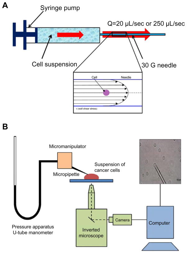
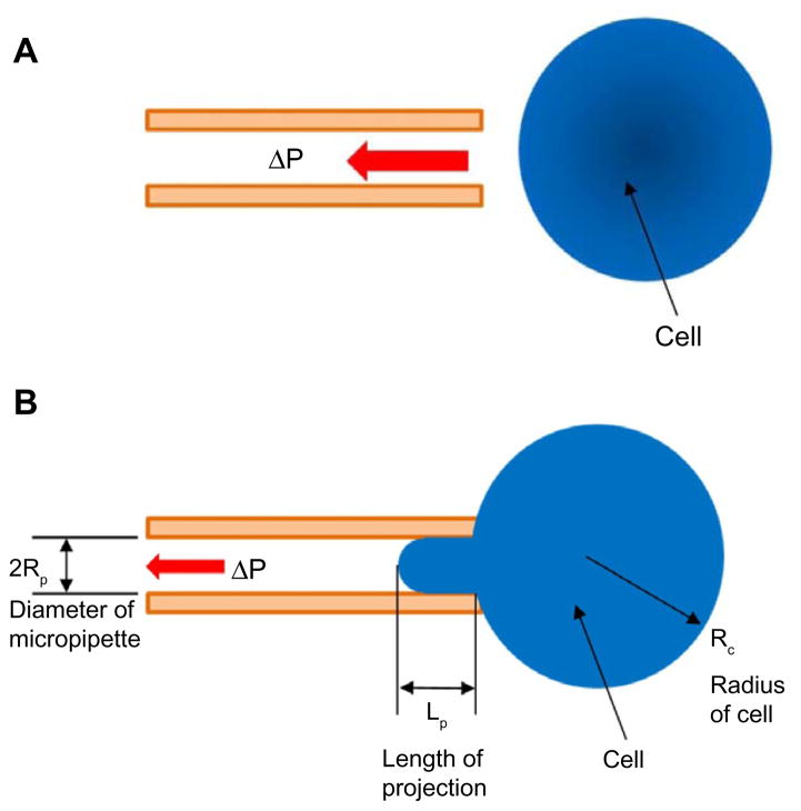
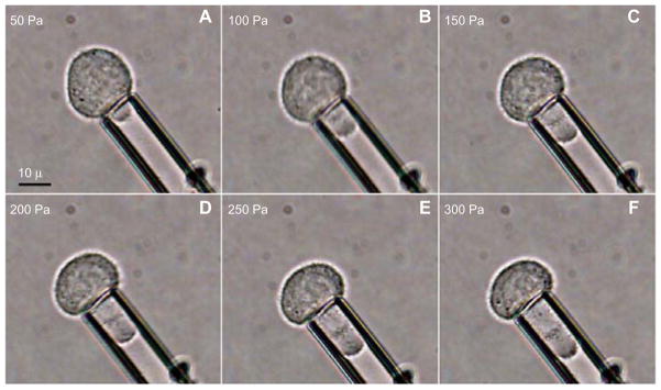
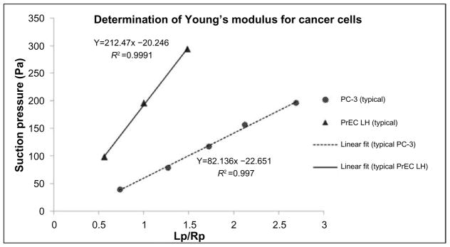
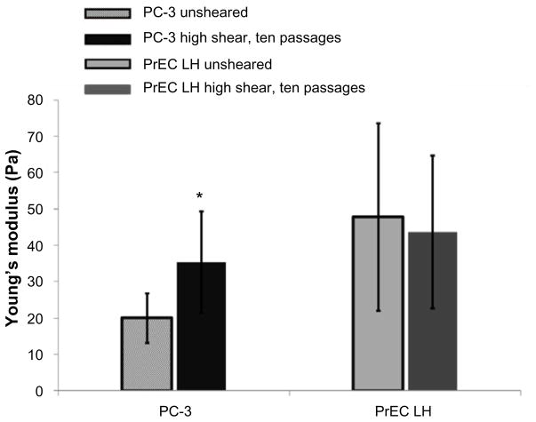
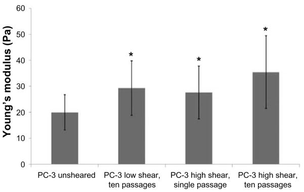
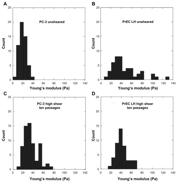
Over 90% of cancer deaths result not from primary tumor development, but from metastatic tumors that arise after cancer cells circulate to distal sites via the circulatory system. While it is known that metastasis is an inefficient process, the effect of hemodynamic parameters such as fluid shear stress (FSS) on the viability and efficacy of metastasis is not well understood. Recent work has shown that select cancer cells may be able to survive and possibly even adapt to FSS in vitro. The current research seeks to characterize the effect of FSS on the mechanical properties of suspended cancer cells in vitro. Nontransformed prostate epithelial cells (PrEC LH) and transformed prostate cancer cells (PC-3) were used in this study. The Young's modulus was determined using micropipette aspiration. We examined cells in suspension but not exposed to FSS (unsheared) and immediately after exposure to high (6,400 dyn/cm2) and low (510 dyn/cm2) FSS. The PrEC LH cells were ~140% stiffer than the PC-3 cells not exposed to FSS. Post-FSS exposure, there was an increase of ~77% in Young's modulus after exposure to high FSS and a ~47% increase in Young's modulus after exposure to low FSS for the PC-3 cells. There was no significant change in the Young's modulus of PrEC LH cells post-FSS exposure. Our findings indicate that cancer cells adapt to FSS, with an increased Young's modulus being one of the adaptive responses, and that this adaptation is specific only to PC-3 cells and is not seen in PrEC LH cells. Moreover, this adaptation appears to be graded in response to the magnitude of FSS experienced by the cancer cells. This is the first study investigating the effect of FSS on the mechanical properties of cancer cells in suspension, and may provide significant insights into the mechanism by which some select cancer cells may survive in the circulation, ultimately leading to metastasis at distal sites. Our findings suggest that biomechanical analysis of cancer cells could aid in identifying and diagnosing cancer in the future.

Keywords: cancer; elastic modulus; fluid shear stress; metastasis; micropipette aspiration.


Conflict of interest statement

Disclosure

The authors report no conflicts of interest in this work.

Figures

Figure 1
Figure 1
Schematic of experimental apparatus to perform fluid shear stress exposure and micropipette aspiration experiments. Notes: (A) Schematic of fluid shear stress exposure apparatus using a syringe pump and 30 G needle, with a schematic of the flow inside the needle shown inset with parabolic velocity profile. (B) Schematic of experimental apparatus for performing micropipette aspiration experiments. The pressure apparatus consists of a fluid reservoir mounted on a linear scale functioning like a u-tube manometer which results in a pressure differential based on raising or lowering of the fluid level in comparison with the level of the microscope stage. The reservoir is connected to an in-house fabricated micropipette mounted on a micromanipulator to manipulate the micropipette toward individual cells. The cell suspension is deposited on a cover slip mounted on an inverted microscope and imaged by a camera connected to a computer. The red and black arrows indicate the direction of flow. Abbreviations: Q, flow rate; sec, second; G, gauge.
Figure 2
Figure 2
Schematic of micropipette aspiration experiment. Notes: (A) A micropipette is manipulated toward a cell and a small suction pressure is applied to attract the cell. (B) The cell is partially aspirated into the micropipette and measurements are taken at different suction pressures, ΔP. Relevant measurements are shown. The red arrows indicate the direction of suction pressure. Abbreviation: ΔP, suction pressure.
Figure 3
Figure 3
Image sequence for a typical micropipette aspiration experiment (AF). Note: The suction pressures are shown on the top left of every image; the images are taken 3–4 minutes apart.
Figure 4
Figure 4
Typical results for relationship of suction pressure and length of projection of the cell inside the pipette. Notes: The length of projection is normalized by the radius of the pipette. The Young’s modulus is obtained from the slope of the linear fits. Abbreviations: PC-3, transformed prostate cancer cells; PrEC LH, immortalized, non-transformed prostate epithelial cells; Lp/Rpm normalized projection length.
Figure 5
Figure 5
Comparison of Young’s modulus for transformed prostate cancer cells (PC-3) and nontransformed prostate epithelial cells (PrEC LH) not exposed to FSS (unsheared) and after exposure to high FSS. Note: There is a ~77% increase in the Young’s modulus of transformed cells after being exposed to high shear, but no discernible change can be seen for the nontransformed cells. *P<0.05. Abbreviation: FSS, fluid shear stress.
Figure 6
Figure 6
Comparison of Young’s modulus for transformed prostate cancer cells (PC-3) not exposed to FSS (unsheared), after exposure to ten passages at high FSS, after exposure to ten passages at low FSS, and after exposure to a single pass at high FSS, respectively. There appears to be a graded response to the level of FSS that the cancer cells are exposed to. *P<0.05. Abbreviation: FSS, fluid shear stress.
Figure 7
Figure 7
Comparison of histogram of Young’s modulus for unexposed (unsheared) and sheared PC-3 and PrEC LH cells (AD). Notes: The different distributions of data can be clearly seen. The standard deviation of the PC-3 unsheared cells is over six times smaller than that of the unsheared PrEC LH cells. Abbreviations: PC-3, transformed prostate cancer cells; PrEC LH, immortalized, non-transformed prostate epithelial cells.

Similar articles

Cited by

References

    1. Wirtz D, Konstantopoulos K, Searson PC. The physics of cancer: the role of physical interactions and mechanical forces in metastasis. Nat Rev Cancer. 2011;11(7):512–522. - PMC - PubMed
    1. Fidler I. The pathogenesis of cancer metastasis: the “seed and soil” hypothesis revisited. Nat Rev Cancer. 2003;3(6):453–458. - PubMed
    1. Mitchell MJ, King MR. Physical biology in cancer. 3. The role of cell glycocalyx in vascular transport of circulating tumor cells. Am J Physiol Cell Physiol. 2014;306(2):C89–C97. - PMC - PubMed
    1. Chaffer CL, Weinberg RA. A perspective on cancer cell metastasis. Science. 2011;331(6024):1559–1564. - PubMed
    1. Kumar S, Weaver VM. Mechanics, malignancy, and metastasis: the force journey of a tumor cell. Cancer Metastasis Rev. 2009;28(1–2):113–127. - PMC - PubMed

LinkOut - more resources