. 2014 Oct 27:5:38.
doi: 10.1186/2041-9139-5-38. eCollection 2014.

Toll signals regulate dorsal-ventral patterning and anterior-posterior placement of the embryo in the hemipteran Rhodnius prolixus

Affiliations

Toll signals regulate dorsal-ventral patterning and anterior-posterior placement of the embryo in the hemipteran Rhodnius prolixus

Mateus Berni et al. Evodevo. .

Abstract

Background: Insect embryonic dorso-ventral patterning depends greatly on two pathways: the Toll pathway and the Bone Morphogenetic Protein pathway. While the relative contribution of each pathway has been investigated in holometabolous insects, their role has not been explored in insects with a hemimetabolous type of development. The hemimetabolous insect Rhodnius prolixus, an important vector of Chagas disease in the Americas, develops from an intermediate germ band and displays complex movements during katatrepsis that are not observed in other orders. However, little is known about the molecular events that regulate its embryogenesis. Here we investigate the expression and function of genes potentially involved in the initial patterning events that establish the embryonic dorso-ventral axis in this hemipteran.

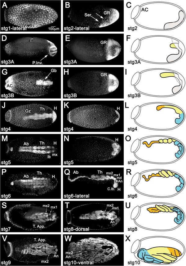
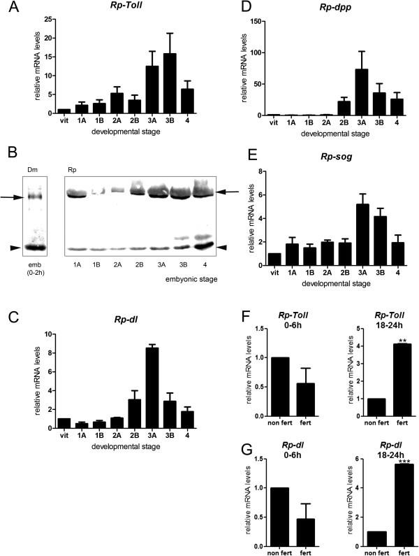
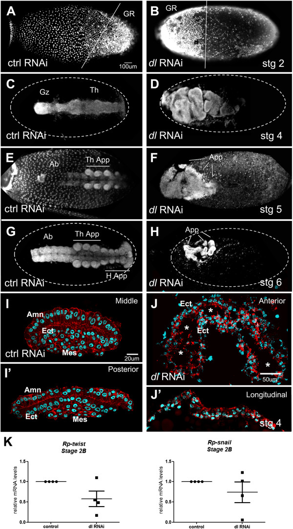
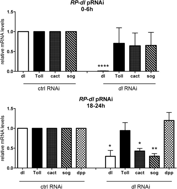
Results: We establish a staging system for early embryogenesis that allows us to correlate embryo morphology with gene expression profiles. Using this system, we investigate the role of Toll pathway genes during embryogenesis. Detailed analyses of gene expression throughout development, coupled with functional analyses using parental RNA interference, revealed that maternal Toll is required to establish germ layers along the dorso-ventral axis and for embryo placement along the anterior-posterior axis. Interestingly, knockdown of the Toll pathway effector Rp-dorsal appears to regulate the expression of the Bone Morphogenetic Protein antagonist Rp-short-gastrulation.

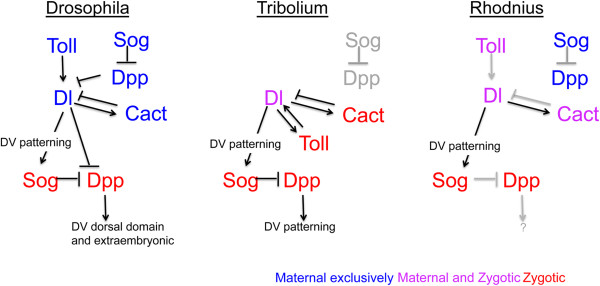
Conclusions: Our results indicate that Toll signals are the initiating event in dorso-ventral patterning during Rhodnius embryogenesis, and this is the first report of a conserved role for Toll in a hemipteran. Furthermore, as Rp-dorsal RNA interference generates anteriorly misplaced embryos, our results indicate a novel role for Toll signals in establishment of the anterior-posterior axis in Rhodnius.

Keywords: Dorso-ventral axis; Embryogenesis; Hemipteran; NFκB; Toll.


Figures

Figure 1
Figure 1
Prospective patterning stages in Rhodnius early embryogenesis. Nuclear staining for fixed embryos at the designated stages. Left and mid panel images are taken from the dorsal and ventral egg surface, respectively, unless stated otherwise. Drawings correspond to graphical representations of the stage in lateral views. (A) Cleavage stage embryo with nuclei at the periphery. (B,C) As the blastoderm develops, nuclei concentrate posteriorly (arrows) to form the germ rudiment (GR, grey in C). Serosal cells (Ser) are placed dorso-anteriorly. (D-F) During gastrulation, posterior invagination (P.Inv) of the GR takes places dorsally and (G-I) the germ band (Gb) extends anteriorly, while on the ventral surface the non-invaginated surface GR (grey in F) forms a horseshoe. Dashed lines in (F) and (I) correspond to the tissue under which the Gb invaginates. (J-L) Once gastrulation is complete, the head (H) and thorax (Th) regions are visible, as well as a growth zone (Gz) from which abdominal structures develop. (M-O) The embryo has grown to its full length. H and Th are segmented, but not the abdominal region (Ab). (P-R) Segmentation is complete. Ab is segmented. The abdominal caudal flexure is immersed in the yolk in (M-R) and consequently not visualized in superior optical sections. (S,T,V) Growth stages in dorsal views. Cuticle fluorescence adds to and takes over nuclear staining with time. (S) Thoracic appendages (T. app.) larger than head appendages. Maxilar appendages mx1 and mx2 are distinguished. (T,U) T. app. curved over ventral surface of the embryo. Mx1 and antennal appendages (An) grow. (V) Mx1 are hidden by An. Throughout the above stages the DV and AP axes of the embryo are inverted respective to the egg. (W,X) During katatrepsis the embryo moves backwards. As a result the embryo and egg axes coincide. Throughout stage 10 the embryo also undergoes dorsal closure. Egg anterior cap (AC); md: mandibule; C. lb.: cephalic lobes. Magnification Bar in (A) is valid for all panels.
Figure 2
Figure 2
Rp-Toll-like-2 is related to other Toll involved in dorso-ventral patterning. (A) Phylogenetic tree illustrating the relationship between Rp Tolls and other Toll-like receptors. (B) Predicted protein structure for Rp-Toll-like-2 (Rp-Toll), compared to Dm-Toll and Tc-Toll, with region recognized by anti-Toll d300 (α-Toll). Sequences used for phylogenetic tree reconstruction were: Nvit-Toll-1A: XP_001604577.1; Nvit-Toll-1B: XP_001604871.2; Nvit-Toll-1C: XP_001604880.2; Nvit-Toll-1D: XP_003425965.1; Dmel-Toll: CG5490-PC, Dmel-Toll2: AAF57509.1, Dmel-Toll-3: AAF86229.1, Dmel-Toll-4: CG18241, Dmel-Toll-5: AAF86227.1, Dmel-Toll-6: CG7250, Dmel-Toll-7: CG8595-PA, Dmel-Toll-8: AAF862241, Dmel-Toll-9: CG5528, Tcas-04901: XP_972312.1, Tc-00176: XP_967154.1, Tcas-04438: XP_967796.1, Tcas-04439: XP_967716.2, Tcas-04452: XP_973926.2, Tcas-04474 XM_967316.2 Tcas-04895: XP_971999.1, Tcas-04898: XP_008200897.1, Tcas-04901: XP_972312.1|, Rpro-Toll-like-1 [VB: RPRC07390], Rpro-Toll like-2 [VB: RPRC 009262], Rpro-Toll-like-3 [VB: RPRC015296], Rpro-Toll like-4 [VB: RPRC004104], Rpro-Toll-like-5: GL562958 (a contig, not a predicted transcript), Rpro-Toll-like-6 [VB: RPRC001608-RA], Agam-Toll1: AAL37901.1, Aaeg-Toll1A: AAEL000057, Aaeg-Toll1B: XP_001658507.1, Amel-Toll: XP_006562783.1 (Nvit: Nasonia vitripennis; Tcas: Tribolium castaneum; Dmel: Drosophila melanogaster; Rpro: Rhodnius prolixus; Agam: Anopheles gambia; Isca: Ixodes scaputari; Aaeg: Aedes aegypti). Genes functionally analyzed for a role in dorso-ventral patterning are displayed in bold, Rp-Toll-like-2 (Rp-Toll) functionally analyzed in this study is in red.
Figure 3
Figure 3
Toll and Bone Morphogenetic Protein pathway genes are dynamically expressed throughout embryonic development. Normalized mRNA levels for (A,F) Rp-Toll, (C,G) Rp-dl, (D) Rp-dpp, and (E) Rp-sog in vitellogenic ovaries (vitelo), embryonic stages (stages 1 to 4, 0 to 48 hours), or unfertilized eggs (F,G). Differences in gene expression throughout embryonic development were significant for all genes between preblastoderm (0 to 12 hours) and gastrulation stages (18 to 30 hours) (one-way analysis of variance, p <0.05). (B) Toll protein levels vary throughout Rhodnius development. (F,G) Differences in mRNA levels for (F) Rp-Toll and (G) Rp-dl in fertilized (fert) versus unfertilized (non fert) 0- to 6-hour or 18- to 24-hour eggs were significant: **p <0.01, ***p <0.001 by paired t-test. Use of appropriate reference genes was defined as in Additional file 1. Dm, D. melanogaster; Rp, R. prolixus.
Figure 4
Figure 4
Toll protein is maternally delivered to developing oocytes. (A) Rhodnius oogenesis showing the tropharium (B), early (C), and late (D) oocytes in vitelarium. Chorionic stages are not displayed. (B) Toll protein is present in follicle cells of the tropharium. (C) Toll in early oocytes is likely delivered from nurse cells through trophic cords. (D) Protein in late oocyte cytoplasm and plasma membrane. Toll protein in green, actin to reveal cell perimeter in red, nuclei DAPI stain in blue. Nc: nurse cells; Oo: oocyte; Tc: trophic cord.
Figure 5
Figure 5
Rp-cact expression and knockdown phenotypes. (A) Normalized mRNA levels for Rp-cact show small variability throughout embryonic development. (B) Rp-cact mRNAs in stage 1A embryos (0 to 6 hours) are mostly maternally provided, while gastrulation stage 2B (18 to 24 hours) mRNAs are generated zygotically, since levels of the former are unaltered, while the latter are decreased in unfertilized eggs (non fert). (C) Rp-cact is also expressed in the immune tissues of the midgut and fat body. (D) In immune tissues such as the gut, Rp-cact levels respond to activation of the Toll pathway using zymosan. (E,F) Ovaries resulting from control MalE (E) or Rp-cact (F) pRNAi show that egg chambers do not develop in the Rp-cact knockdown.
Figure 6
Figure 6
Rp-dl knockdown embryonic phenotypes. Embryos resulting from control (A,C,E,G,I) or Rp-dl (B,D,F,H,J) parental RNA interference (pRNAi). (A,B) At the blastoderm stage control (ctrl) embryos present a posteriorly localized germ rudiment (GR) that is diagonally displayed (A, as in Figure  2B), while the Rp-dl RNAi GR is anteriorly localized and perpendicular to the egg’s long axis. (C,D) After gastrulation the head, thorax (Th), and growth zone (Gz) are seen in control (C) MalE RNAi embryos, but are not distinguished in (D) the Rp-dl RNAi. (E,F) Stage 5 and (G,H) stage 6 embryos show the correct formation of appendages in control (E,G) MalE RNAi embryos, while (F,H) Rp-dl RNAi embryos present only appendage-like structures (App.) that are seen in 25% of cases. These structures cannot be identified as head (H.App.) or thoracic appendages (Th. App.). Ab, abdomen. (I-J’) After gastrulation, amnion (Amn), ectodermal (Ect) and mesodermal (Mes) layers are distinguished in control embryo transverse sections stained with Alexa 647-phalloidin and nuclear Hoescht (I,I’). Shown are embryo sections in the middle (I) and posterior (I’) regions of the egg. Rp-dl RNAi (J,J’) embryos placed at the egg anterior form a hollow tube (Ect) with no distinguishable mesodermal layer, as seen in transverse (J) and longitudinal (J’) sections. Note that these embryos are localized adjacent to the egg surface. The asterisks in J point to the hollow embryo interior. Lateral view of embryos in (A,B; A slightly tilted ventrally), dorsal view in (C-H). However, embryo dorso-ventral placement in Rp-dl RNAi is randomized. (K) Relative mRNA levels for mesodermal genes in control and Rp-dl RNAi embryos (four biological replicates). Rp-twist and Rp-snail show a tendency to decrease in Rp-dl knockdowns.
Figure 7
Figure 7
Rp-sog and Rp-cact mRNA levels are regulated by Rp-dl . Normalized mRNA levels for Rp-dl, Rp-Toll, Rp-cact, Rp-sog, and Rp-dpp in control mal parental RNA interference (pRNAi) or Rp-dl pRNAi (0.5 μg/μL) embryos were evaluated at stage 1 (0 to 6 hours) and stage 2B (18 to 24 hours) embryonic development. *p <0.05, **p <0.01, ****p < 0.0001 by paired t-test.
Figure 8
Figure 8
Model for the conserved action of Toll signals during Rhodnius dorso-ventral patterning. The hierarchical relationships between Toll and Bone Morphogenetic Protein pathways in dorso-ventral (DV) patterning have been established for Drosophila and Tribolium, either in terms of gene transcription or protein activity, and are displayed in simplified form to compare with relationships defined here for Rhodnius. Neither Tribolium nor Rhodnius Toll pathways regulate dpp expression, activity required to pattern the dorsal region of the Drosophila embryo. On the other hand, a feedback loop between Toll and dl is only present in Tribolium. In the three species analyzed, dl positively regulates sog expression. Arrows in black are those tested experimentally. Arrows in grey denote relations that are assumed based on conserved aspects and published evidence throughout the animal kingdom. Maternal, zygotic or maternal + zygotic expression are displayed in blue, red and pink, respectively. Maternal (blue) and zygotic (red) expression of dpp and sog are not gathered as one in Drosophila and Rhodnius since their epistatic relationships to the Toll pathway differ (Drosophila), or have not been fully analyzed (Rhodnius).

References

    1. Davis GK, Patel NH. Short, long, and beyond: molecular and embryological approaches to insect segmentation. Annu Rev Entomol. 2002;47:669–699. doi: 10.1146/annurev.ento.47.091201.145251. - DOI - PubMed
    1. Lynch JA, Roth S. The evolution of dorsal-ventral patterning mechanisms in insects. Genes Dev. 2011;25:107–118. doi: 10.1101/gad.2010711. - DOI - PMC - PubMed
    1. Moussian B, Roth S. Dorsoventral axis formation in the Drosophila embryo - shaping and transducing a morphogen gradient. Curr Biol. 2005;15:R887–R899. doi: 10.1016/j.cub.2005.10.026. - DOI - PubMed
    1. Chen G, Handel K, Roth S. The maternal NF-kappaB/dorsal gradient of Tribolium castaneum: dynamics of early dorsoventral patterning in a short-germ beetle. Development. 2000;127:5145–5156. - PubMed
    1. Nunes da Fonseca R R, von Levetzow C, Kalscheuer P, Basal A, van der Zee M, Roth S. Self-regulatory circuits in dorsoventral axis formation of the short-germ beetle Tribolium castaneum. Dev Cell. 2008;14:605–615. doi: 10.1016/j.devcel.2008.02.011. - DOI - PubMed