. 2015 Mar 9;6(4):1172-94.
doi: 10.1364/BOE.6.001172. eCollection 2015 Apr 1.

Kernel regression based segmentation of optical coherence tomography images with diabetic macular edema

Affiliations

Kernel regression based segmentation of optical coherence tomography images with diabetic macular edema

Stephanie J Chiu et al. Biomed Opt Express. .

Abstract

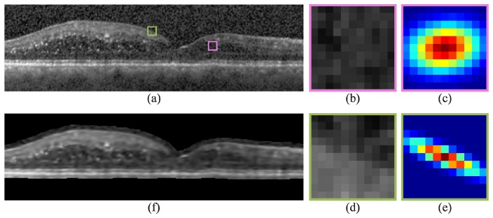
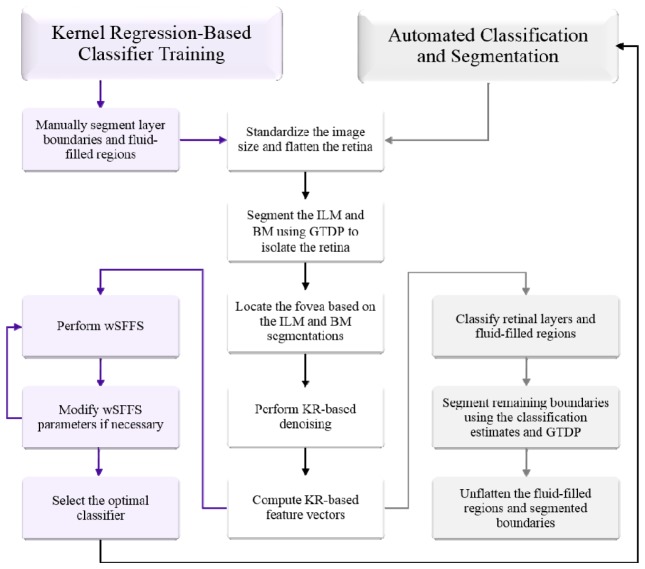
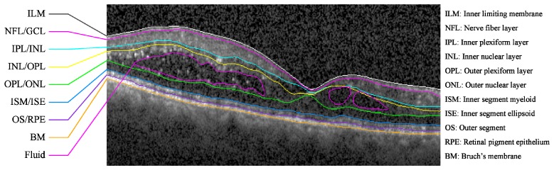
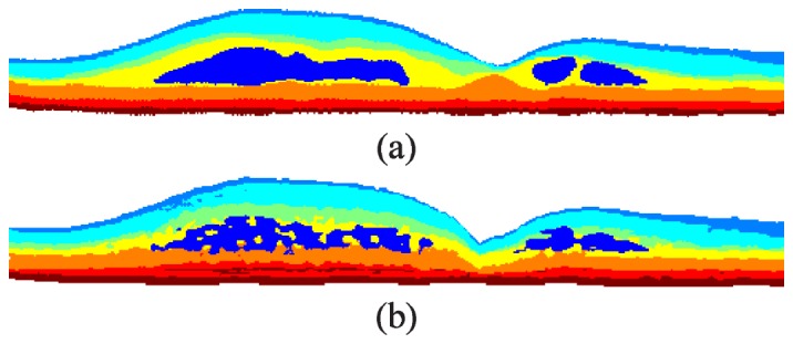
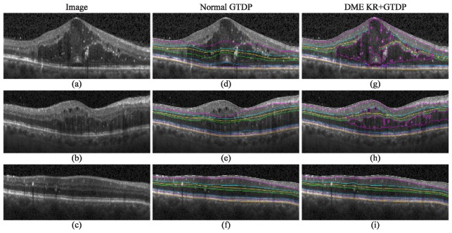
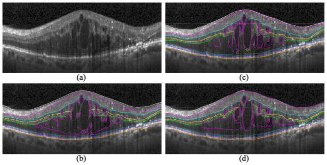
We present a fully automatic algorithm to identify fluid-filled regions and seven retinal layers on spectral domain optical coherence tomography images of eyes with diabetic macular edema (DME). To achieve this, we developed a kernel regression (KR)-based classification method to estimate fluid and retinal layer positions. We then used these classification estimates as a guide to more accurately segment the retinal layer boundaries using our previously described graph theory and dynamic programming (GTDP) framework. We validated our algorithm on 110 B-scans from ten patients with severe DME pathology, showing an overall mean Dice coefficient of 0.78 when comparing our KR + GTDP algorithm to an expert grader. This is comparable to the inter-observer Dice coefficient of 0.79. The entire data set is available online, including our automatic and manual segmentation results. To the best of our knowledge, this is the first validated, fully-automated, seven-layer and fluid segmentation method which has been applied to real-world images containing severe DME.

Keywords: (100.0100) Image processing; (170.4470) Ophthalmology; (170.4500) Optical coherence tomography.


Figures

Fig. 1
Fig. 1
Second order iterative Gaussian steering KR of an SD-OCT image with DME. a) Automatically flattened image, b,d) zoomed-in images of the pink and green boxes in (a), c,e) Gaussian steering kernels used to denoised the central pixel of (b) and (d), and f) KR-denoised image of (a). In (f), regions external to the retina (black) were not denoised with the exception of a padded boundary surrounding the retina required for feature vector computation.
Fig. 2
Fig. 2
Flowchart of the KR-based classification and GTDP-based segmentation algorithm for identifying fluid-filled regions and eight retinal layer boundaries on images with DME pathology.
Fig. 3
Fig. 3
Target retinal layer boundaries and fluid to segment on an SD-OCT B-scan of an eye with DME. A flattened version of the image without markings is shown in Fig. 1(a).
Fig. 4
Fig. 4
KR-based classification of retinal layers and fluid-filled regions. a) Manual classification of the image in Fig. 1(a) with the classes defined in Table 1. b) Automatic classification using the merged DME classifier.
Fig. 5
Fig. 5
Laws’ E5TE5 texture feature computed for the image in Fig. 1(a) with (a) and without (b) KR-based denoising. Both feature images have been enhanced using adaptive histogram equalization.
Fig. 6
Fig. 6
Visual example of features selected for the DME classifiers. a-l) Features computed for the image in Fig. 1(a).
Fig. 7
Fig. 7
Qualitative results for the identification of fluid and eight retinal layer boundaries on SD-OCT images of eyes with DME. a-c) Images with significant, moderate, and no visible fluid, respectively, d-f) their corresponding automatic segmentation result using the GTDP algorithm developed for normal eyes, and g-i) the automatic KR + GTDP segmentation result.
Fig. 8
Fig. 8
Automatic and manual segmentation comparison. a) An SD-OCT B-scan with DME pathology, b) the fully automatic segmentation result, and c-d) the segmentation results completed by two different graders.
Fig. 9
Fig. 9
Automatic fluid detection errors. Top) Cropped portions of SD-OCT B-scans with DME, middle) manual segmentation of fluid-filled regions by a grader, and bottom) automatic fluid-filled classification. a) An image with hyper-reflective deposits, b) a dim image with a hyper-reflective outlier, and c) a high quality image.

References

    1. Davidson J. A., Ciulla T. A., McGill J. B., Kles K. A., Anderson P. W., “How the diabetic eye loses vision,” Endocrine 32(1), 107–116 (2007). 10.1007/s12020-007-0040-9 - DOI - PubMed
    1. Yau J. W., Rogers S. L., Kawasaki R., Lamoureux E. L., Kowalski J. W., Bek T., Chen S. J., Dekker J. M., Fletcher A., Grauslund J., Haffner S., Hamman R. F., Ikram M. K., Kayama T., Klein B. E., Klein R., Krishnaiah S., Mayurasakorn K., O’Hare J. P., Orchard T. J., Porta M., Rema M., Roy M. S., Sharma T., Shaw J., Taylor H., Tielsch J. M., Varma R., Wang J. J., Wang N., West S., Xu L., Yasuda M., Zhang X., Mitchell P., Wong T. Y., Meta-Analysis for Eye Disease (META-EYE) Study Group , “Global prevalence and major risk factors of diabetic retinopathy,” Diabetes Care 35(3), 556–564 (2012). 10.2337/dc11-1909 - DOI - PMC - PubMed
    1. Kumar V., Abbas A. K., Fausto N., Robbins S. L., Cotran R. S., Robbins and Cotran pathologic basis of disease, 7th ed. (Elsevier Saunders, Philadelphia, 2005), pp. xv, 1525 p.
    1. Pe’er J., Folberg R., Itin A., Gnessin H., Hemo I., Keshet E., “Upregulated expression of vascular endothelial growth factor in proliferative diabetic retinopathy,” Br. J. Ophthalmol. 80(3), 241–245 (1996). 10.1136/bjo.80.3.241 - DOI - PMC - PubMed
    1. Tolentino M. J., McLeod D. S., Taomoto M., Otsuji T., Adamis A. P., Lutty G. A., “Pathologic features of vascular endothelial growth factor-induced retinopathy in the nonhuman primate,” Am. J. Ophthalmol. 133(3), 373–385 (2002). 10.1016/S0002-9394(01)01381-2 - DOI - PubMed

LinkOut - more resources