. 2015 Mar 25;6(4):1487-501.
doi: 10.1364/BOE.6.001487. eCollection 2015 Apr 1.

Measuring the optical characteristics of medulloblastoma with optical coherence tomography

Affiliations

Measuring the optical characteristics of medulloblastoma with optical coherence tomography

Barry Vuong et al. Biomed Opt Express. .

Abstract

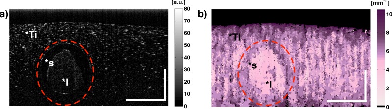
Medulloblastoma is the most common malignant pediatric brain tumor. Standard treatment consists of surgical resection, followed by radiation and high-dose chemotherapy. Despite these efforts, recurrence is common, leading to reduced patient survival. Even with successful treatment, there are often severe long-term neurologic impacts on the developing nervous system. We present two quantitative techniques that use a high-resolution optical imaging modality: optical coherence tomography (OCT) to measure refractive index, and the optical attenuation coefficient. To the best of our knowledge, this study is the first to demonstrate OCT analysis of medulloblastoma. Refractive index and optical attenuation coefficient were able to differentiate between normal brain tissue and medulloblastoma in mouse models. More specifically, optical attenuation coefficient imaging of normal cerebellum displayed layers of grey matter and white matter, which were indistinguishable in the structural OCT image. The morphology of the tumor was distinct in the optical attenuation coefficient imaging. These inherent properties may be useful during neurosurgical intervention to better delineate tumor boundaries and minimize resection of normal tissue.

Keywords: (100.2960) Image analysis; (110.4500) Optical coherence tomography; (170.6935) Tissue characterization; (290.1350) Backscattering.


Figures

Fig. 1
Fig. 1
(a) An OCT image of human fingertips. (b) The result of mean shift segmentation from the OCT image based on clustering. Each color in the mean shift segmentation image represents a segmented area. Scale bar is 1mm.
Fig. 2
Fig. 2
(a) Gross histology of harvested Math1-GFP; Ptch+/− mouse model. (b) The corresponding fluorescence image. (c) The combined gross and fluorescence image. (d) A 500 μm thick section of the fluorescent medulloblastoma (red dashed box) region was imaged by OCT on a planar reflective surface. Refractive index was measured by evaluating the optical thickness of the specimen (z + z′). Scale bar is 1 mm.
Fig. 3
Fig. 3
(a) Structural OCT image of a cortical phantom which consisted of titanium dioxide versa gel (*Ti), silicon tube (*s) and diluted intralipid (*I). (b) The corresponding OCT-OA image of the cortical phantom. Scale bar: 1 mm.
Fig. 4
Fig. 4
(a) Gross histology of harvested Math1-GFP; Ptch+/− mouse model. (b) The corresponding fluorescence image. (c) The combined gross and fluorescence image. Normal cerebellum (cyan line) and an interface of normal/medulloblastoma (red line) scans were taken. (d) H&E section of the cerebellum. Grey matter consisting of molecular layer (*M) and granular layer (*G) was observed. Underneath the granular layer, regions of white matter are present (*W). (e) The associated structural OCT with minimal resolved features. (f) The corresponding OCT-OA image consisted of a layered structure. The shape and location of these layers are similar to the molecular layer (*M) and granular layer (*G). Scale bar 500 μm
Fig. 5
Fig. 5
(a) H&E section of the cerebellum in a Math1-GFP; Ptch+/− model. Grey matter consisting of molecular layer (*M) and granular layer (*G) was observed. Underneath the granular layer, regions of white matter (*W) are present. Regions of medulloblastoma resided on the right side (*T). Within the red dashed box, medulloblastoma regions that were adjacent to normal cerebellum demonstrated very dense cellular proliferation (red arrow). On the right side of the medulloblastoma region, superficial cells appear to be sparsely spaced (green arrow); however a lining of dense cells was located below. (b) The corresponding structural OCT with minimal resolved features. (c) The OCT-OA image consisted of a layered structure and erratic high attenuation region. In normal tissue the layers consisted of the molecular layer (*M) and granular layer (*G). The erratic high attenuation region was where medulloblastoma (*T). Scale bar 500 μm.
Fig. 6
Fig. 6
(a) H&E section of a normal cerebellum where the molecular layer (*M) and granular layer (*G) are visualized. (b) The associated structural OCT image. (c) The corresponding OCT-OA image consisted of a high optical attenuation coefficient region (granular) and low optical attenuation (molecular). Scale bar 1 mm.
Fig. 7
Fig. 7
(a) A surface prospective (transverse plane) from 3D reconstruction of structural OCT. Normal cerebellum (*C) and medulloblastoma (*T) regions were difficult to differentiate. (b) The same perspective was rendered from 3D OCT-OA images. Regions of normal cerebellum and medulloblastoma are clearly visualized. Both 3D OCT and 3D OCT-OA consisted of the visualization of a superficial blood vessel (white arrows). (c) Cross sectional regions in the 3D structural OCT image demonstrated few morphological features. (d) The corresponding 3D OCT-OA image exhibits layered structures. In the cross sectional regions, key morphological features were observed. The molecular layer (green arrow), granular layer (black arrow) and white matter (red arrow) could be resolved. A fly-through of the entire 3D OCT-OA is shown in Media 1. Scale bar 1 mm.

Similar articles

Cited by

References

    1. Gilbertson R. J., “Medulloblastoma: signalling a change in treatment,” The Lancet Oncology 5, 209–218 (2004).10.1016/S1470-2045(04)01424-X - DOI - PubMed
    1. Legler J. M., Ries L. A. G., Smith M. A., Warren J. L., Heineman E. F., Kaplan R. S., Linet M. S., “Brain and other central nervous system cancers: recent trends in incidence and mortality,” J. Nat. Cancer Inst. 91, 1382–1390 (1999).10.1093/jnci/91.16.1382 - DOI - PubMed
    1. Gajjar A., Hernan R., Kocak M., Fuller C., Lee Y., McKinnon P. J., Wallace D., Lau C., Chintagumpala M., Ashley D. M., Kellie S. J., Kun L., Gilbertson R. J., “Clinical, histopathologic, and molecular markers of prognosis: toward a new disease risk stratification system for medulloblastoma,” J. Clin. Oncol. 22, 984–993 (2004).10.1200/JCO.2004.06.032 - DOI - PubMed
    1. Kumar V., Abbas A. K., Fausto N., Aster J. C., Robbins and Cotran Pathologic Basis of Disease, Professional Edition: Expert Consult-Online, 8 (Elsevier Health Sciences, 2009).
    1. Pui C.-H., Gajjar A. J., Kane J. R., Qaddoumi I. A., Pappo A. S., “Challenging issues in pediatric oncology,” Nat. Rev. Clin. Oncol. 8, 540–549 (2011).10.1038/nrclinonc.2011.95 - DOI - PMC - PubMed

LinkOut - more resources