Dissociated multimodal hubs and seizures in temporal lobe epilepsy
- PMID: 25909080
- PMCID: PMC4402080
- DOI: 10.1002/acn3.173
Dissociated multimodal hubs and seizures in temporal lobe epilepsy
Abstract
Objective: Brain connectivity at rest is altered in temporal lobe epilepsy (TLE), particularly in "hub" areas such as the posterior default mode network (DMN). Although both functional and anatomical connectivity are disturbed in TLE, the relationships between measures as well as to seizure frequency remain unclear. We aim to clarify these associations using connectivity measures specifically sensitive to hubs.
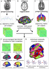
Methods: Connectivity between 1000 cortical surface parcels was determined in 49 TLE patients and 23 controls with diffusion and resting-state functional magnetic resonance imaging. Two types of hub connectivity were investigated across multiple brain modules (the DMN, motor system, etcetera): (1) within-module connectivity (a measure of local importance that assesses a parcel's communication level within its own subnetwork) and (2) between-module connectivity (a measure that assesses connections across multiple modules).
Results: In TLE patients, there was lower overall functional integrity of the DMN as well as an increase in posterior hub connections with other modules. Anatomical between-module connectivity was globally decreased. Higher DMN disintegration (DD) coincided with higher anatomical between-module connectivity, whereas both were associated with increased seizure frequency. DD related to seizure frequency through mediating effects of anatomical connectivity, but seizure frequency also correlated with anatomical connectivity through DD, indicating a complex interaction between multimodal networks and symptoms.
Interpretation: We provide evidence for dissociated anatomical and functional hub connectivity in TLE. Moreover, shifts in functional hub connections from within to outside the DMN, an overall loss of integrative anatomical communication, and the interaction between the two increase seizure frequency.
Figures


Similar articles
-
Increased Intrinsic Connectivity of the Default Mode Network in Temporal Lobe Epilepsy: Evidence from Resting-State MEG Recordings.PLoS One. 2015 Jun 2;10(6):e0128787. doi: 10.1371/journal.pone.0128787. eCollection 2015. PLoS One. 2015. PMID: 26035750 Free PMC article.
-
Association between seizure freedom and default mode network reorganization in patients with unilateral temporal lobe epilepsy.Epilepsy Behav. 2019 Jan;90:238-246. doi: 10.1016/j.yebeh.2018.10.025. Epub 2018 Dec 8. Epilepsy Behav. 2019. PMID: 30538081
-
Parcellation of the Hippocampus Using Resting Functional Connectivity in Temporal Lobe Epilepsy.Front Neurol. 2019 Aug 22;10:920. doi: 10.3389/fneur.2019.00920. eCollection 2019. Front Neurol. 2019. PMID: 31507522 Free PMC article.
-
The structural and functional connectivity of the posterior cingulate cortex: comparison between deterministic and probabilistic tractography for the investigation of structure-function relationships.Neuroimage. 2014 Nov 15;102 Pt 1:118-27. doi: 10.1016/j.neuroimage.2013.12.022. Epub 2013 Dec 21. Neuroimage. 2014. PMID: 24365673 Review.
-
Hub overload and failure as a final common pathway in neurological brain network disorders.Netw Neurosci. 2024 Apr 1;8(1):1-23. doi: 10.1162/netn_a_00339. eCollection 2024. Netw Neurosci. 2024. PMID: 38562292 Free PMC article. Review.
Cited by
-
MRI characterization of temporal lobe epilepsy using rapidly measurable spatial indices with hemisphere asymmetries and gender features.Neuroradiology. 2015 Sep;57(9):873-86. doi: 10.1007/s00234-015-1540-6. Epub 2015 Jun 2. Neuroradiology. 2015. PMID: 26032924
-
Presurgical Mapping of the Language Network Using Resting-state Functional Connectivity.Top Magn Reson Imaging. 2016 Feb;25(1):19-24. doi: 10.1097/RMR.0000000000000073. Top Magn Reson Imaging. 2016. PMID: 26848557 Free PMC article. Review.
-
A Mapping Between Structural and Functional Brain Networks.Brain Connect. 2016 May;6(4):298-311. doi: 10.1089/brain.2015.0408. Epub 2016 Mar 29. Brain Connect. 2016. PMID: 26860437 Free PMC article.
-
Postoperative seizure freedom does not normalize altered connectivity in temporal lobe epilepsy.Epilepsia. 2017 Nov;58(11):1842-1851. doi: 10.1111/epi.13867. Epub 2017 Aug 3. Epilepsia. 2017. PMID: 28776646 Free PMC article.
-
Structural covariance networks relate to the severity of epilepsy with focal-onset seizures.Neuroimage Clin. 2018;20:861-867. doi: 10.1016/j.nicl.2018.09.023. Epub 2018 Sep 26. Neuroimage Clin. 2018. PMID: 30278373 Free PMC article.
References
-
- Gusnard DA, Raichle ME. Searching for a baseline: functional imaging and the resting human brain. Nat Rev Neurosci. 2001;2:685–694. - PubMed
-
- Van Diessen E, Diederen SJH, Braun KPJ, et al. Functional and structural brain networks in epilepsy: what have we learned? Epilepsia. 2013;54:1855–1865. - PubMed
Grants and funding
LinkOut - more resources
Full Text Sources
Other Literature Sources

