. 2015 Apr;2(4):362-72.
doi: 10.1002/acn3.179. Epub 2015 Feb 16.

Endplate denervation correlates with Nogo-A muscle expression in amyotrophic lateral sclerosis patients

Affiliations

Endplate denervation correlates with Nogo-A muscle expression in amyotrophic lateral sclerosis patients

Gaëlle Bruneteau et al. Ann Clin Transl Neurol. 2015 Apr.

Abstract

Objective: Data from mouse models of amyotrophic lateral sclerosis (ALS) suggest early morphological changes in neuromuscular junctions (NMJs), with loss of nerve-muscle contact. Overexpression of the neurite outgrowth inhibitor Nogo-A in muscle may play a role in this loss of endplate innervation.

Methods: We used confocal and electron microscopy to study the structure of the NMJs in muscle samples collected from nine ALS patients (five early-stage patients and four long-term survivors). We correlated the morphological results with clinical and electrophysiological data, and with Nogo-A muscle expression level.

Results: Surface electromyography assessment of neuromuscular transmission was abnormal in 3/9 ALS patients. The postsynaptic apparatus was morphologically altered for almost all NMJs (n = 430) analyzed using confocal microscopy. 19.7% of the NMJs were completely denervated (fragmented synaptic gutters and absence of nerve terminal profile). The terminal axonal arborization was usually sparsely branched and 56.8% of innervated NMJs showed a typical reinnervation pattern. Terminal Schwann cell (TSC) morphology was altered with extensive cytoplasmic processes. A marked intrusion of TSCs in the synaptic cleft was seen in some cases, strikingly reducing the synaptic surface available for neuromuscular transmission. Finally, high-level expression of Nogo-A in muscle was significantly associated with higher extent of NMJ denervation and negative functional outcome.

Interpretation: Our results support the hypothesis that morphological alterations of NMJs are present from early-stage disease and may significantly contribute to functional motor impairment in ALS patients. Muscle expression of Nogo-A is associated with NMJ denervation and thus constitutes a therapeutic target to slow disease progression.


Figures
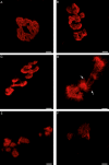
Figure 1
Figure 1
Morphology of the postsynaptic apparatus observed by confocal microscopy (α-Bungarotoxin staining of AChRs, in red). (A) Normal, “pretzel-shaped,” postsynaptic apparatus. (B–E) Denervation-induced modifications of the postsynaptic apparatus. Fragmentation and enlargement of the postsynaptic apparatus with flattening of the primary synaptic gutter. The outer edge of the postsynaptic primary gutter sometimes showed spike-like areas (D, arrows). (F) Barely visible postsynaptic apparatus, usually seen in the absence of contact with a nerve terminal profile, that is, completely denervated NMJ. Scale bars, 5 μm. AChR, acetylcholine receptor; NMJ, neuromuscular junction.
Figure 2
Figure 2
Ultrastructural findings at the neuromuscular junction (NMJ). (A) Control and (B–E) patients with amyotrophic lateral sclerosis (ALS). (A) Normal NMJ from a control subject. The primary synaptic gutter is occupied by a nerve terminal (N) containing numerous synaptic vesicles and mitochondria. Note the postsynaptic membrane folding with well-formed secondary synaptic clefts (arrow). The terminal Schwann cell (TSC) caps the nerve terminal, without extending into the synaptic cleft (the asterisk is placed over the TSC nucleus). (B) A flattened primary synaptic gutter with well-preserved subneural folds (arrow) faces a remodeled motor nerve terminal. The vacuoles within the nerve ending (N) are probably artifacts. The TSC caps the nerve terminal (star: TSC nucleus). Multiple layers of TSC basal lamina can be seen near the primary synaptic cleft (arrowheads). (C–E) Denervated NMJs. (C) Partially denervated NMJ. A small nerve terminal profile (N) partially occupies the primary synaptic gutter. Note the well-preserved secondary synaptic clefts (white arrow) and the TSC capping the nerve terminal (arrowheads). The presence of acetylcholine receptors (AChRs) on the crests of the subneural folds is identified as an electron-opaque border (black arrows). (D) Same NMJ as in B, lower magnification. On the crests of the subneural folds, where AChRs are present, the muscle postsynaptic membrane shows an electron-opaque border (arrow). Here, this dark border extends to the immediately adjacent, unfolded, postsynaptic membrane (arrowheads). (E) Fully denervated NMJ. Unoccupied synaptic gutter with absence of nerve terminal and well-preserved subneural folds (arrow). Residual TSC basal lamina can be seen in the remaining primary synaptic cleft (arrowhead). Scale bars, 2 μm.
Figure 3
Figure 3
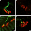
Neuromuscular junctions (NMJs) observed by confocal microscopy. Staining of acetylcholine receptors (AChRs) with α-bungarotoxin, in red, and of axon terminals with neurofilament antibody, in green. (A) Normal NMJ from one control (profile view). The motor axon divides into two first-order branches. (B) Fully denervated NMJ with no axon. Note the low AChR staining of the postsynaptic compartment. (C) Reinnervated NMJ. A fragmented postsynaptic apparatus is innervated by a motor axon which divides into two first-order branches (arrowheads). Note the fine nodal sprout reinnervating two postsynaptic elements (arrow). (D) Remodeled, partially innervated NMJ. The axon terminal partially innervates a slightly flattened postsynaptic apparatus. Scale bars, 5 μm.
Figure 4
Figure 4
Neuromuscular junctions (NMJs) observed by confocal microscopy after staining of acetylcholine receptors (AChRs) with α-bungarotoxin, in red, and of terminal Schwann cells (TSCs) with anti-S100 antibody, in green. (A) A TSC (arrow) caps a postsynaptic apparatus made of two adjacent synaptic cups. There are no Schwann cell cytoplasmic processes. (B) A TSC (arrow) elaborates two cytoplasmic processes (arrowheads), each process contacting one isolated postsynaptic element. Scale bars, 5 μm.
Figure 5
Figure 5
Electron microscopy view showing an interposition of the terminal Schwann cell (TSC) between the axon terminal and the postsynaptic muscle membrane. (A, B) The membrane of the TSC (star) invades the synaptic cleft (arrowhead) and encases the nerve terminal (N). Secondary synaptic clefts are maintained (white arrow). Note in (B) that the area of the muscle postsynaptic membrane facing the residual part of the nerve terminal (black arrow), available for neurotransmission, is very narrow. Scale bar, 2 μm.
Figure 6
Figure 6
High-level expression of Nogo-A in muscle is associated with a faster functional decline and a higher proportion of denervated neuromuscular junctions (NMJs). Nogo-A muscle protein level, determined by the optical density of the Nogo-A signal bands normalized to the Red Ponceau staining (a.u.: arbitrary units), correlated negatively with the disease duration, and positively with the disease progression rate and the proportion of denervated NMJs. (A) Negative correlation between muscle Nogo-A protein content and disease duration at the time of the biopsy (Spearman rank correlation r = −0.893). (B) Positive correlation between muscle Nogo-A protein level and the disease progression rate from symptom onset (Spearman rank correlation r = 0.893). (C) Positive correlation between muscle Nogo-A protein level and the proportion of denervated NMJs (Spearman rank correlation r = 0.821).

References

    1. Charcot JM, Joffroy A. Deux cas d'atrophie musculaire progressive avec lésions de la substance grise et des faisceaux antéro-latéraux de la moelle épinière. Arch Physiol. 1869;2:354–367.
    1. Beghi E, Mennini T, Bendotti C, et al. The heterogeneity of amyotrophic lateral sclerosis: a possible explanation of treatment failure. Curr Med Chem. 2007;14:3185–3200. - PubMed
    1. Fischer LR, Culver DG, Tennant P, et al. Amyotrophic lateral sclerosis is a distal axonopathy: evidence in mice and man. Exp Neurol. 2004;185:232–240. - PubMed
    1. Vinsant S, Mansfield C, Jimenez-Moreno R, et al. Characterization of early pathogenesis in the SOD1(G93A) mouse model of ALS: Part II, results and discussion. Brain Behav. 2013;3:431–457. - PMC - PubMed
    1. Frey D, Schneider C, Xu L, et al. Early and selective loss of neuromuscular synapse subtypes with low sprouting competence in motoneuron diseases. J Neurosci. 2000;20:2534–2542. - PMC - PubMed