. 2015 Apr;2(4):388-400.
doi: 10.1002/acn3.182. Epub 2015 Feb 19.

Blocking GluR2-GAPDH ameliorates experimental autoimmune encephalomyelitis

Affiliations

Blocking GluR2-GAPDH ameliorates experimental autoimmune encephalomyelitis

Dongxu Zhai et al. Ann Clin Transl Neurol. 2015 Apr.

Abstract

Objective: Multiple sclerosis (MS) is the most common disabling neurological disease of young adults. The pathophysiological mechanism of MS remains largely unknown and no cure is available. Current clinical treatments for MS modulate the immune system, with the rationale that autoimmunity is at the core of MS pathophysiology.

Methods: Experimental autoimmune encephalitis (EAE) was induced in mice with MOG35-55 and clinical scoring was performed to monitor signs of paralysis. EAE mice were injected intraperitoneally with TAT-fusion peptides daily from day 10 until day 30 after immunization, and their effects were measured at day 17 or day 30.

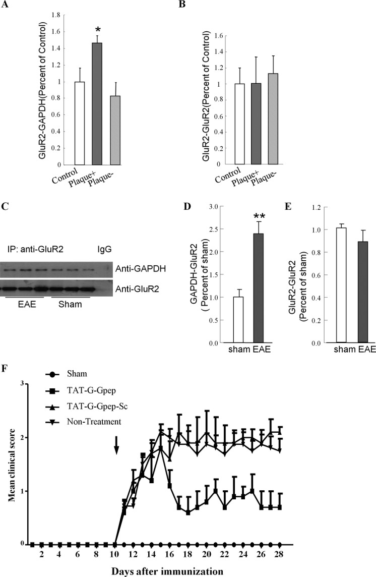
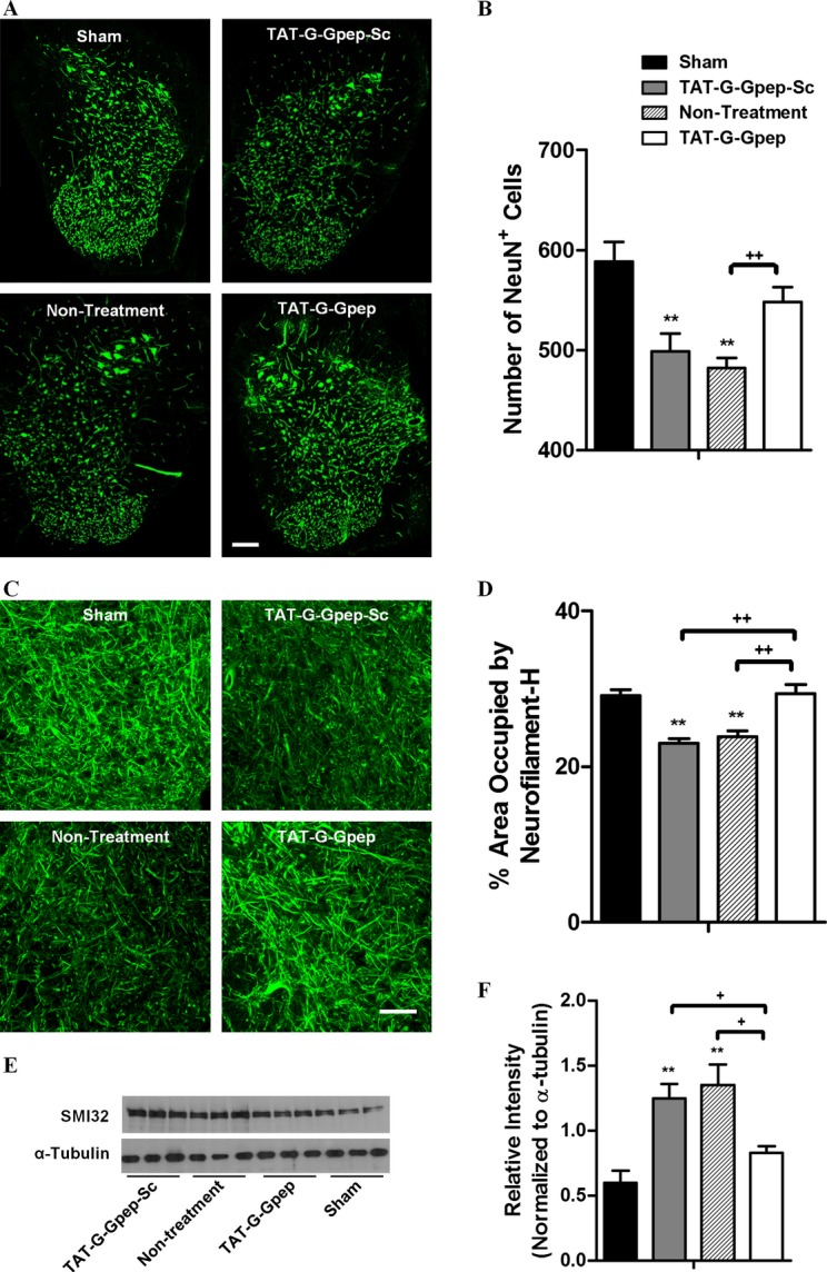
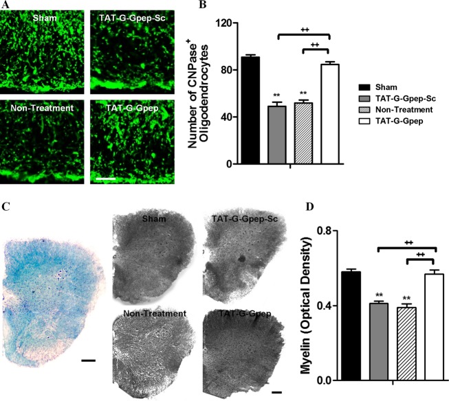
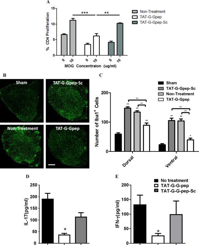
Results: We report a novel target for the development of MS therapy, which aimed at blocking glutamate-mediated neurotoxicity through targeting the interaction between the AMPA (2-amino-3-(3-hydroxy-5-methyl-isoxazol-4-yl) propanoic acid) receptor and an interacting protein. We found that protein complex composed of the GluR2 subunit of AMPA receptors and GAPDH (glyceraldehyde-3-phosphate dehydrogenase) was present at significantly higher levels in postmortem tissue from MS patients and in EAE mice, an animal model for MS. Next, we developed a peptide that specifically disrupts the GluR2 -GAPDH complex. This peptide greatly improves neurological function in EAE mice, reduces neuron death, rescues demyelination, increases oligodendrocyte survival, and reduces axonal damage in the spinal cords of EAE mice. More importantly, our peptide has no direct suppressive effect on naive T-cell responses or basal neurotransmission.

Interpretation: The GluR2 -GAPDH complex represents a novel therapeutic target for the development of medications for MS that work through a different mechanism than existing treatments.


Figures

Figure 1
Figure 1
GluR2–GAPDH complex in multiple sclerosis (MS). (A, B) GluR2–GAPDH complex formation is significantly increased in the plaque of multiple sclerosis (MS). Postmortem samples from control, MS plaque (plaque+) area, and MS nonplaque (plaque−) area were incubated with GluR2 antibody and the precipitated proteins were immunoblotted with GAPDH antibody or GluR2 antibody. The intensity of each protein band for GAPDH (A), GluR2 (B) from all three groups was quantified by densitometry (AIS software, Imaging Research Inc.). Results for each sample are presented as the percentage of the mean of the control samples on the same blot. (*< 0.05, n = 8, one-way ANOVA). (C–E) GluR2–GAPDH complex formation is significantly increased in experimental autoimmune encephalitis (EAE) mice compared to sham mice. (A) Representative image of western blot analysis of GAPDH (top) and GluR2 (bottom) levels precipitated by GluR2 antibody in extract prepared from mouse spinal cord tissue were incubated with GluR2 antibody. Precipitated proteins were subject to SDS-PAGE (sodium dodecyl sulfate polyacrylamide gel electrophoresis) and immunoblotted with GAPDH or GluR2 antibody. (D, E) Densitometric analysis of the level of GAPDH and GluR2. The intensity of GAPDH and GluR2 was quantified by densitometry (software: Image J, NIH). Data were analyzed by t-test. (**< 0.01, n = 3). (F) Clinical EAE scores (mean ± SEM) over time of four groups vaccinated with MOG35-55 on day 0 and treated intraperitoneally daily with TAT-G-Gpep and TAT-G-Gpep-Sc from day 10 (arrow). Starting from day 12, < 0.05, data were analyzed by Mann–Whitney U test.
Figure 2
Figure 2
TAT-G-Gpep treatment rescues both neuronal and axonal density in mouse spinal cords with experimental autoimmune encephalitis (EAE). (A) Representative images of mouse spinal cords immunostained against NeuN+ in sham, TAT-G-Gpep-Sc, nontreatment and TAT-G-Gpep treatment groups. Scale bar = 100 μm. (B) There was a significantly fewer total number of neurons in Tat-G-Gpep-Sc and nontreatment groups when compared to sham animals. However, TAT-G-Gpep treatment resulted in a significant increase in NeuN+ cells versus nontreatment mice, and was comparable to sham controls. There was also an increasing trend observed with TAT-G-Gpep treatment when compared to TAT-G-Gpep-Sc group. (C) Neurofilament-H immunostained images of mouse spinal cords in sham, TAT-G-Gpep-Sc, nontreatment and TAT-G-Gpep treatment groups. Scale bar = 15 μm. (D) Quantification of neurofilament-H staining was converted to a black and white threshold scale and expressed as a percentage of area occupied by fluorescence in the dorsal and ventral funiculi of mouse spinal cords. The area occupied by neurofilament-H labeling was significantly lower in TAT-G-Gpep-Sc and nontreated mice when compared to sham, while TAT-G-Gpep-treated mice showed a significant rescue in axon density which was comparable to sham controls. All data are shown as mean ± SEM; *< 0.05, **< 0.01 versus sham; ++< 0.01. (E) Axonal damage, assessed by western blot for abnormally dephosphorylated neurofilament-H. Western blot analysis of whole spinal cord homogenate, visualized by enhanced chemiluminescent ECL, of proteins from all four groups: sham, TAT-G-Gpep-Sc, nontreatment and TAT-G-Gpep treatment groups. (F) Densitometric analysis of western blots of total spinal cord homogenate of three representative mice per group, developed by horseradish peroxidase/3, 3'-diaminobenzidine HRP/DAB. Data represent means ± SEM. Differences between groups were accessed by Student–Newman–Keuls post hoc one-way ANOVA **< 0.01 versus sham controls, +< 0.05.
Figure 3
Figure 3
Administration of TAT-G-Gpep significantly promotes oligodendrocyte survival and rescues demyelination in the experimental autoimmune encephalitis (EAE) mouse spinal cords. (A) Representative images of fluorescently labeled CNPase-reactive oligodendrocytes in sham, TAT-G-Gpep-Sc, nontreated, and TAT-G-Gpep-treated mouse spinal cords. Scale bar = 15 μm. (B) There were significantly fewer CNPase-immunolabeled oligodendrocytes in TAT-G-Gpep-Sc and nontreated mice when compared to sham. But TAT-G-Gpep treatment significantly rescued oligodendrocyte numbers to a level comparable to sham controls. (C) Representative Luxol Fast Blue image of an EAE mouse spinal cord section with no treatment is shown in blue. Luxol Fast Blue staining was used to quantify myelination (blue) in different groups. Color images converted to gray scale values of the four groups are shown on the right. Scale bar = 100 μm. (D) Myelin density was measured as optical density according to precalibrated values in black and white transmission. Similarly, significant demyelination was observed in TAT-G-Gpep-Sc and nontreated mice when compared to sham. TAT-G-Gpep treatment significantly increased myelination to sham control levels. All data are shown as mean ± SEM; **< 0.01 versus sham, ++< 0.01.
Figure 4
Figure 4
TAT-G-Gpep treatment diminishes the activated immune response in experimental autoimmune encephalitis (EAE) mice. (A) EAE mice in all groups showed an increase in CD4+ T-cell proliferation when presented with 10 μg/mL of MOG. TAT-G-Gpep administration significantly reduced this proliferative response. **< 0.01; ***< 0.001. (B) Representative images of Iba1-immunolabeled macrophages/microglia in sham, TAT-G-Gpep-Sc, nontreated and TAT-G-Gpep-treated mouse spinal cords. Scale bar = 100 μm. (C) Quantification of the number of Iba1+ cells in the dorsal and ventral horns revealed significantly more macrophages/microglia residing in scrambled peptide, nontreated and peptide-treated mice when compared to sham. Peptide treatment significantly reduced the amount Iba1+ cells when compared to scrambled peptide or nontreated mice. All data are shown as mean ± SEM; **< 0.01 versus sham; +< 0.01. (D, E) TAT-G-Gpep treatment in EAE mice resulted in a significant reduction in IL-17 (D) and IFN-γ (E) levels when compared to nontreated or TAT-G-Gpep-Sc treated mice. *< 0.05.

Similar articles

Cited by

References

    1. Trapp BD, Stys PK. Virtual hypoxia and chronic necrosis of demyelinated axons in multiple sclerosis. Lancet Neurol. 2009;8:280–291. - PubMed
    1. Compston A, Coles A. Multiple sclerosis. Lancet. 2008;372:1502–1517. - PubMed
    1. Kieseier BC, Wiendl H, Hemmer B, Hartung HP. Treatment and treatment trials in multiple sclerosis. Curr Opin Neurol. 2007;20:286–293. - PubMed
    1. Trapp BD, Nave KA. Multiple sclerosis: an immune or neurodegenerative disorder? Annu Rev Neurosci. 2008;31:247–269. - PubMed
    1. Stys PK, Zamponi GW, van Minnen J, Geurts JJ. Will the real multiple sclerosis please stand up? Nat Rev Neurosci. 2012;13:507–514. - PubMed