. 2015 Apr 24;10(4):e0123934.
doi: 10.1371/journal.pone.0123934. eCollection 2015.

Structural Analysis of the 14-3-3ζ/Chibby Interaction Involved in Wnt/β-Catenin Signaling

Affiliations

Structural Analysis of the 14-3-3ζ/Chibby Interaction Involved in Wnt/β-Catenin Signaling

Ryan C Killoran et al. PLoS One. .

Abstract

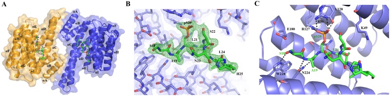
The partially disordered Chibby (Cby) is a conserved nuclear protein that antagonizes the Wnt/β-catenin signaling pathway. By competing with the Tcf/Lef family proteins for binding to β-catenin, Cby abrogates the β-catenin-mediated transcription of Wnt signaling genes. Additionally, upon phosphorylation on S20 by the kinase Akt, Cby forms a complex with 14-3-3 to facilitate the nuclear export of β-catenin, which represents another crucial mechanism for the regulation of Wnt signaling. To obtain a mechanistic understanding of the 14-3-3/Cby interaction, we have extensively characterized the complex using X-ray crystallography, nuclear magnetic resonance (NMR) spectroscopy, and isothermal titration calorimetry (ITC). The crystal structure of the human 14-3-3ζ/Cby protein-peptide complex reveals a canonical binding mode; however the residue at the +2 position from the phosphorylated serine is shown to be uniquely oriented relative to other solved structures of 14-3-3 complexes. Our ITC results illustrate that although the phosphorylation of S20 is essential for Cby to recognize 14-3-3, residues flanking the phosphorylation site also contribute to the binding affinity. However, as is commonly observed in other 14-3-3/phosphopeptide crystal structures, residues of Cby flanking the 14-3-3 binding motif lack observable electron density. To obtain a more detailed binding interface, we have completed the backbone NMR resonance assignment of 14-3-3ζ. NMR titration experiments reveal that residues outside of the 14-3-3 conserved binding cleft, namely a flexible loop consisting of residues 203-210, are also involved in binding Cby. By using a combined X-ray and NMR approach, we have dissected the molecular basis of the 14-3-3/Cby interaction.


Conflict of interest statement

Competing Interests: The authors have declared that no competing interests exist.

Figures

Fig 1
Fig 1
(A) Crystal structure of the 14-3-3ζ /Cby 18-mer complex. Each 14-3-3ζ monomer (coloured orange and blue) is bound to one Cby phosphopeptide. (B) Final 2F oF c electron density of 14-3-3ζ (blue mesh) and Cby 18-mer (green mesh) contoured to 1σ. (C) Polar contacts (black hashed lines) between 14-3-3ζ residues (light blue sticks) and the Cby 18-mer (green sticks).
Fig 2
Fig 2. Structural comparison of 14-3-3ζ-bound Cby with other 14-3-3 binding motifs comprising various +2 residues.
Residues K49 and R56 are coloured yellow on the white surface representation of 14-3-3ζ. Structural and sequence alignment of Cby (green sticks) with the binding-motifs of (A). Raf1 (pink sticks, PDB: 3CU8), (B) PKCε (orange sticks, PDB: 2WH0), (C) Histone H3 (white sticks, PDB: 2C1N), (D) β2 integrin (blue sticks, PDB: 2V7D) and (E) α 4 integrin (purple sticks. PDB: 4HKC). The Cα RMSD values were computed by subtracting a Cα distance matrix between the -2 and +1 residues of Cby and each peptide as well as the -2 and most C-terminal residue in the respective alignments.
Fig 3
Fig 3
(A) 1H-15N TROSY-HSQC spectrum and backbone resonance assignment of 2H/13C/15N labeled 14-3-3ζΔC12. (B) Secondary structure propensity (SSP) scores for 14-3-3ζΔC12. SSP scores were calculated based upon the 13Cα/β chemical shifts.
Fig 4
Fig 4. NMR titration experiments of 14-3-3ζΔC12 with Cby peptides.
(A) 1H-15N TROSY-HSQC spectra of 14-3-3ζΔC12 alone (black) and with 3 molar equivalents of the Cby 7-mer (red) and Cby 18-mer (blue). (B) 1H-15N TROSY-HSQC spectra of 14-3-3ζΔC12 alone (black) and with 3 molar equivalents of the WT Cby 18-mer (blue) and Cby S22P 18-mer (blue).
Fig 5
Fig 5. The mapping of chemical shifts on the crystal structure of the 14-3-3ζ /Cby complex.
Due to the crowding of some peaks, the chemical shifts of some residues could not be confidently traced and were excluded from the analysis. (A) (Above) A monomer of the 14-3-3ζ /Cby crystal structure interface based on Cby residues (18SApSLSNLH25). Residues R56, R127 and Y128 which contact the Cby phosphate group are coloured red, residues interfacing the peptide are coloured cyan, and 14-3-3ζ residues coloured magenta are found along the 14-3-3 dimer interface. (Below) The 14-3-3ζ dimer bound to Cby. (B) (Cby 7-mer), (C) (Cby 18-mer) and (D) (Cby S22P 18-mer). Residues with traceable assigned resonances are coloured on a blue—white—yellow gradient (0 ppm to 0.1 ppm) based on their combined chemical shift [Δω = ((Δδ1H)2 +(0.2*Δδ15N)2)1/2] at a 3:1 peptide:protein ratio. Residues coloured in orange represent peaks that broadened out to disappearance upon addition of peptide. Residues R56, R127 and Y128 are coloured pink.
Fig 6
Fig 6. The 14-3-3ζ, Cby, β-catenin tripartite complex.
Model of dimeric 14-3-3ζ bound to two molecules of full-length Cby. The models of two full-length Cby proteins have disordered residues 30–73 of the N-terminus and 100–126 of the C-terminus presented in a highly extended fashion, and are shown forming a coiled-coil from residues 73–100. β-catenin (PDB: 2Z6H) is shown with its Cby binding-site (helix C) in red which then binds along the C-terminus (64–126) of Cby.

Similar articles

Cited by

References

    1. Takemaru K, Yamaguchi S, Lee YS, Zhang Y, Carthew RW, Moon RT (2003) Chibby, a nuclear beta-catenin-associated antagonist of the Wnt/Wingless pathway. Nature 422: 905–909. - PubMed
    1. Clevers H (2006) Wnt/beta-catenin signaling in development and disease. Cell 127: 469–480. - PubMed
    1. Logan CY, Nusse R (2004) The Wnt signaling pathway in development and disease. Annu Rev Cell Dev Biol 20: 781–810. - PubMed
    1. Kahn M (2014) Can we safely target the WNT pathway? Nat Rev Drug Discov 13: 513–532. 10.1038/nrd4233 - DOI - PMC - PubMed
    1. Thakur R, Mishra DP (2013) Pharmacological modulation of beta-catenin and its applications in cancer therapy. J Cell Mol Med 17: 449–456. 10.1111/jcmm.12033 - DOI - PMC - PubMed

Publication types

MeSH terms