

High USP22 expression indicates poor prognosis in hepatocellular carcinoma
- PMID: 25909224
- PMCID: PMC4494964
- DOI: 10.18632/oncotarget.3705
High USP22 expression indicates poor prognosis in hepatocellular carcinoma
Abstract
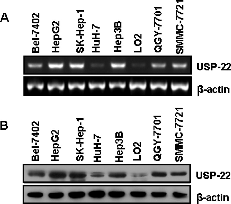
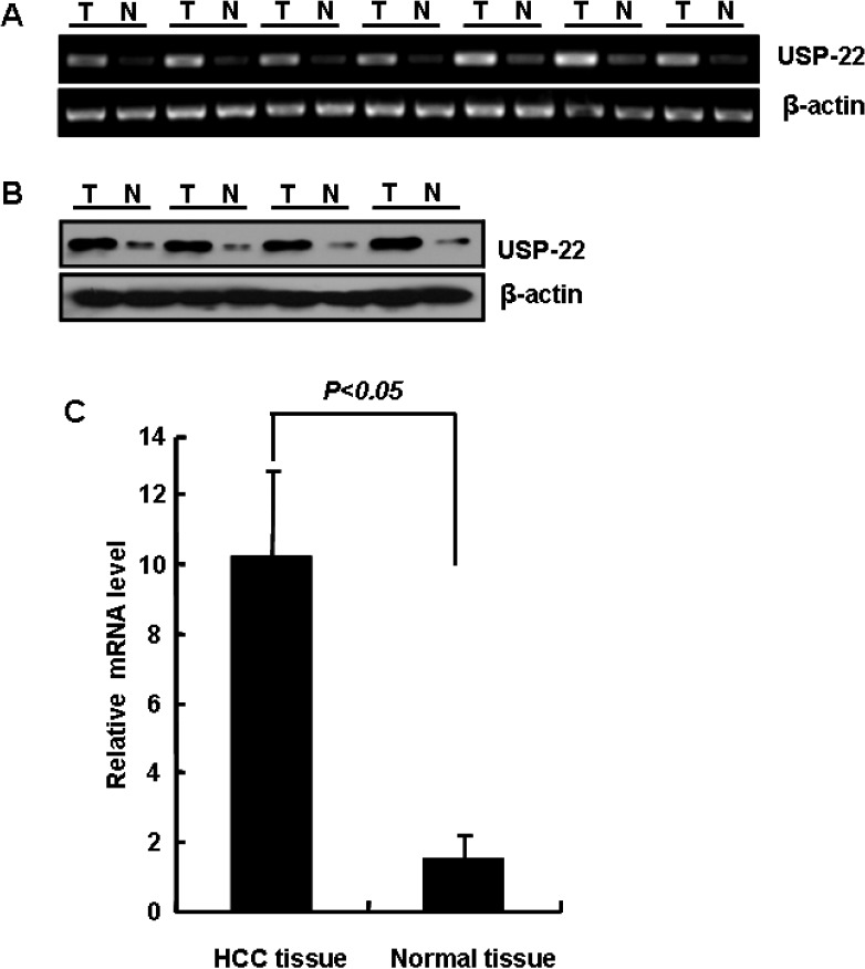
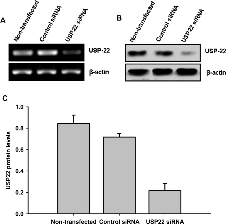
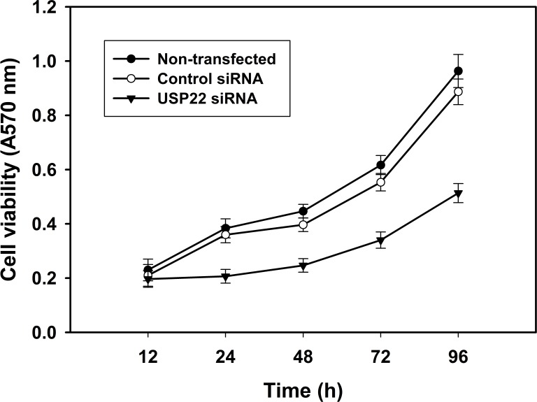
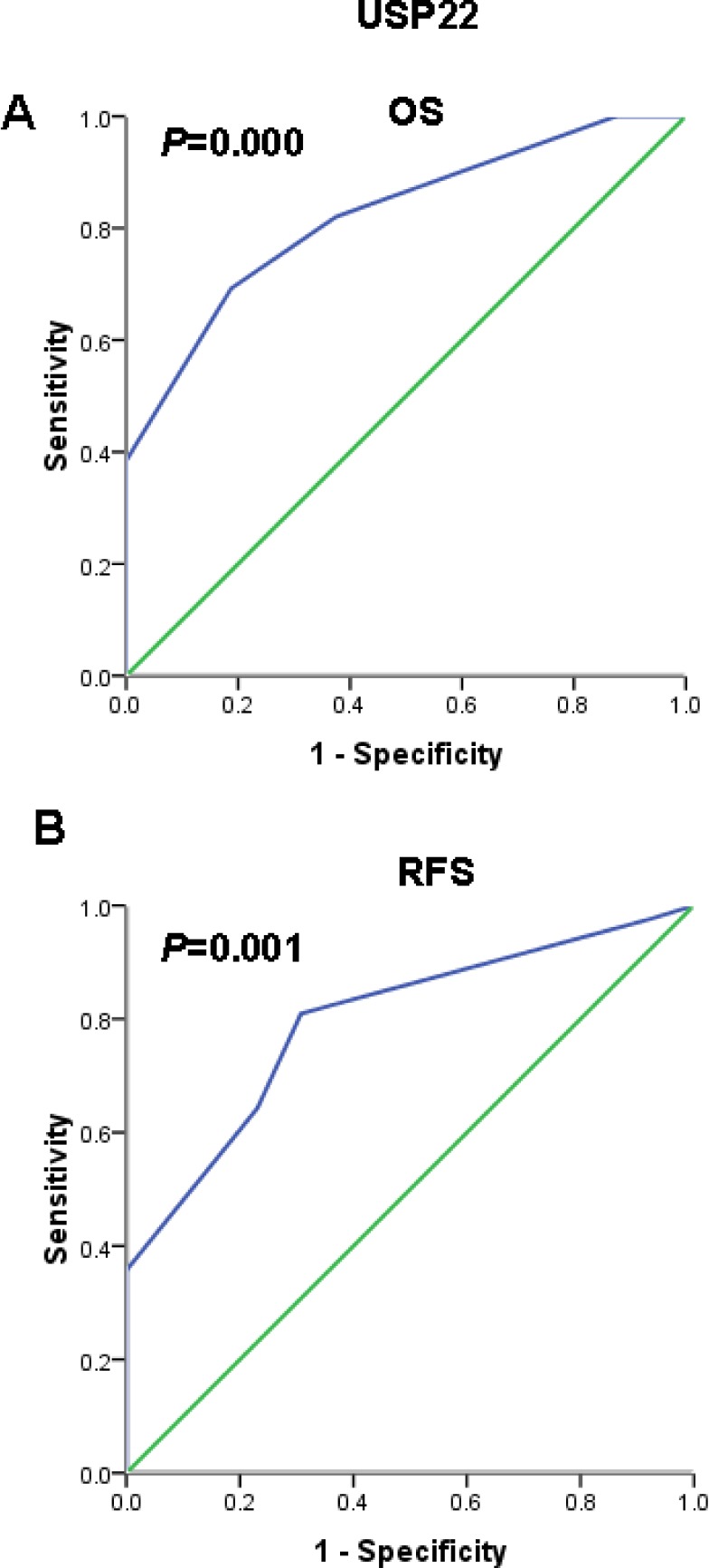
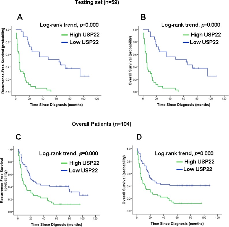
Ubiquitin-specific protease 22 (USP22) removes ubiquitin from histones, thus regulating gene transcription. The expression frequency and expression levels of USP22 were significantly higher in hepatocellular carcinoma (HCC) than in normal liver tissues. High USP22 expression in HCC was significantly correlated with clinical stage and tumor grade. Kaplan-Meier analysis showed that elevated USP22 expression predicted poorer overall survival and recurrence-free survival. High USP22 expression was also associated with shortened survival time in patients at advanced tumor stages and with high grade HCC. Multivariate analyses revealed that USP22 expression is an independent prognostic parameter in HCC. These findings provide evidence that high USP22 expression might be important in tumor progression and serves as an independent molecular marker for poor HCC prognosis. Thus, USP22 overexpression identifies patients at high risk and represents a novel therapeutic molecular target for this tumor.
Keywords: cancer biomarker; hepatocellular carcinoma; prognosis; ubiquitin-specific protease 22.
Conflict of interest statement
All authors declare no conflict of interest.
Figures
Similar articles
-
Expression of USP22 and Survivin is an indicator of malignant behavior in hepatocellular carcinoma.Int J Oncol. 2015 Dec;47(6):2208-16. doi: 10.3892/ijo.2015.3214. Epub 2015 Oct 20. Int J Oncol. 2015. Retraction in: Int J Oncol. 2023 Mar;62(3):37. doi: 10.3892/ijo.2023.5485. PMID: 26497847 Retracted.
-
Decreased H2B monoubiquitination and overexpression of ubiquitin-specific protease enzyme 22 in malignant colon carcinoma.Hum Pathol. 2015 Jul;46(7):1006-14. doi: 10.1016/j.humpath.2015.04.001. Epub 2015 Apr 15. Hum Pathol. 2015. PMID: 25971547
-
Neuron-glial antigen 2 overexpression in hepatocellular carcinoma predicts poor prognosis.World J Gastroenterol. 2015 Jun 7;21(21):6649-59. doi: 10.3748/wjg.v21.i21.6649. World J Gastroenterol. 2015. PMID: 26074703 Free PMC article.
-
Ubiquitin Specific Peptidase 22 Regulates Histone H2B Mono-Ubiquitination and Exhibits Both Oncogenic and Tumor Suppressor Roles in Cancer.Cancers (Basel). 2017 Dec 6;9(12):167. doi: 10.3390/cancers9120167. Cancers (Basel). 2017. PMID: 29210986 Free PMC article. Review.
-
Ubiquitin-specific peptidase 22 functions and its involvement in disease.Oncotarget. 2016 Jul 12;7(28):44848-44856. doi: 10.18632/oncotarget.8602. Oncotarget. 2016. PMID: 27057639 Free PMC article. Review.
Cited by
-
USP22 Functions as an Oncogenic Driver in Prostate Cancer by Regulating Cell Proliferation and DNA Repair.Cancer Res. 2020 Feb 1;80(3):430-443. doi: 10.1158/0008-5472.CAN-19-1033. Epub 2019 Nov 18. Cancer Res. 2020. PMID: 31740444 Free PMC article.
-
Ubiquitin-Specific Protease 22/Silent Information Regulator 1 Axis Plays a Pivotal Role in the Prognosis and 5-Fluorouracil Resistance in Hepatocellular Carcinoma.Dig Dis Sci. 2020 Apr;65(4):1064-1073. doi: 10.1007/s10620-019-05844-8. Epub 2019 Oct 5. Dig Dis Sci. 2020. PMID: 31587155 Free PMC article.
-
Immune Evasion and Drug Resistance Mediated by USP22 in Cancer: Novel Targets and Mechanisms.Front Immunol. 2022 Jul 20;13:918314. doi: 10.3389/fimmu.2022.918314. eCollection 2022. Front Immunol. 2022. PMID: 35935969 Free PMC article. Review.
-
Pirarubicin reduces USP22 expression by inhibiting CREB-1 phosphorylation in HeLa cells.Exp Ther Med. 2019 May;17(5):4230-4236. doi: 10.3892/etm.2019.7447. Epub 2019 Mar 27. Exp Ther Med. 2019. PMID: 31007754 Free PMC article.
-
Upregulation of USP22 and ABCC1 during Sorafenib Treatment of Hepatocellular Carcinoma Contribute to Development of Resistance.Cells. 2022 Feb 11;11(4):634. doi: 10.3390/cells11040634. Cells. 2022. PMID: 35203285 Free PMC article.
References
-
- Parkin DM, Bray F, Ferlay J, Pisani P. Global cancer statistics, 2002. CA Cancer J Clin. 2005;55:74–108. - PubMed
-
- Bosch FX, Ribes J, Díaz M, Cléries R. Primary liver cancer: worldwide incidence and trends. Gastroenterology. 2004;127:S5–S16. - PubMed
-
- Lee HJ, Kim MS, Shin JM, Park TJ, Chung HM, Baek KH. The expression patatterns of deubiquitinating enzymes, USP22 and Usp22. Gene Expr Patterns. 2006;6:277–284. - PubMed
Publication types
MeSH terms
Substances
LinkOut - more resources
Full Text Sources
Other Literature Sources
Medical
Research Materials
