. 2015 Jun 2;54(21):3269-85.
doi: 10.1021/acs.biochem.5b00012. Epub 2015 May 21.

Structural features of a 3' splice site in influenza a

Affiliations

Structural features of a 3' splice site in influenza a

Jonathan L Chen et al. Biochemistry. .

Abstract

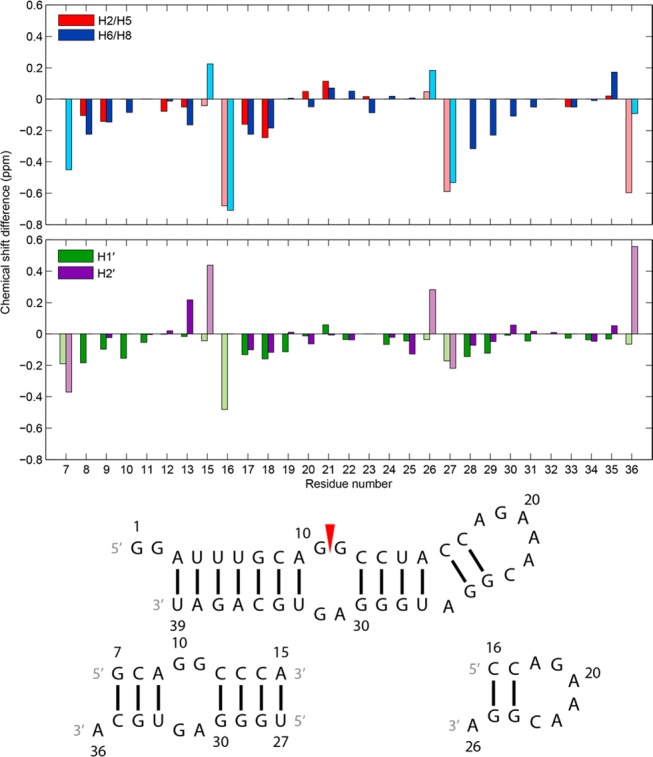
Influenza A is an RNA virus with a genome of eight negative sense segments. Segment 7 mRNA contains a 3' splice site for alternative splicing to encode the essential M2 protein. On the basis of sequence alignment and chemical mapping experiments, the secondary structure surrounding the 3' splice site has an internal loop, adenine bulge, and hairpin loop when it is in the hairpin conformation that exposes the 3' splice site. We report structural features of a three-dimensional model of the hairpin derived from nuclear magnetic resonance spectra and simulated annealing with restrained molecular dynamics. Additional insight was provided by modeling based on (1)H chemical shifts. The internal loop containing the 3' splice site has a dynamic guanosine and a stable imino (cis Watson-Crick/Watson-Crick) GA pair. The adenine bulge also appears to be dynamic with the A either stacked in the stem or forming a base triple with a Watson-Crick GC pair. The hairpin loop is a GAAA tetraloop closed by an AC pair.


Figures

Figure 1
Figure 1
Secondary structures of constructs of the 3′ splice site region of segment 7 mRNA. A red arrowhead denotes the splice site. (a) Pseudoknot and hairpin conformations from ref (15). The SF2/ASF exonic splicing enhancer binding site is colored green and a polypyrimidine tract blue. Numbers in brackets correspond to numbering of residues of the 39 nt hairpin studied with NMR. (b) The 39 nt hairpin studied with NMR. (c) The 11 nt hairpin mimic. (d) The 19 nt duplex model containing the 2 nt × 2 nt internal loop. The U14 residue in the 39-mer was substituted with a cytidine to stabilize formation of the target heterodimer over a homodimer.
Figure 2
Figure 2
Imino proton region of 1D and 2D proton NMR spectra of the 39 nt construct showing sequential proton walks with blue and green lines. The water signal was suppressed with a 1–1–echo pulse in the 1D spectrum and an S-pulse in the 2D NOESY spectrum. The daggers in the 1D spectrum mark chemical exchange peaks. The spectra were acquired at −2 °C with a mixing time of 125 ms for the 2D spectrum. Addition of 5 and 10 mM Mg2+ caused minor shifts and sharpening of the imino resonances, including U4, U5, U6, G11, G19, G24, and U33 (Figure S3 of the Supporting Information).
Figure 3
Figure 3
Schematic of the secondary structure of the 39 nt hairpin with assigned interresidue NOEs. Blue lines denote NOEs identified in the 39-mer and the 19 nt duplex, green lines NOEs identified in the 39-mer and the 11 nt hairpin, and red lines NOEs identified only in the 39-mer.
Figure 4
Figure 4
Differences in chemical shifts between the 39 nt hairpin and 19 nt duplex (residues 7–15 and 27–36) and between the 39 nt hairpin and 11 nt hairpin (residues 16–26) for select nonexchangeable aromatic and sugar protons. Chemical shift data were obtained from spectra at 20 °C for the 39-mer and 11-mer and at 25 °C for the 19-mer. Spectra for the 11-mer and 19-mer were acquired with 5 mM Mg2+. Bars colored with light shades belong to terminal helix residues of the 11-mer (residues 16 and 26) and 19-mer (residues 7, 15, 27, and 36) that are not at the termini of any helices of the 39-mer and thus are in structurally inequivalent regions among the constructs. Residue numbers on the x-axis align with the middle of each set of two bars in each plot. Residue 14 is not included because of a U to C substitution.
Figure 5
Figure 5
H1′–H6/H8 region of a 2D proton NOESY spectrum of the 39 nt hairpin showing sequential proton walks for residues C12–C23 and G24–A31. The C23H1′–H6 and C23H1′–G24H8 NOEs are missing from the walk because the C23H1′ and H2O resonances are close to each other. H1′–H6/H8 walk NOEs are labeled in blue. Adenine H2 signals are labeled with red dashed lines. H1′–adenine H2 NOEs are labeled in red with only the label of the residue for H1′. A G19H1′ (5.45 ppm)–A21H8 (7.96 ppm) NOE is labeled in orange. The spectrum was acquired at 20 °C and a mixing time of 350 ms with a WATERGATE pulse to suppress the water signal. In the secondary structure of the 39 nt hairpin, residues whose intraresidue H1′–H6/H8 NOEs were identified in the NOESY walks are labeled in blue. Spectrum and walks for residues G2−G10 and U33−U39 are in Figure S5 of the Supporting Information.
Figure 6
Figure 6
(a) H1′–H6/H8 region of a 2D proton NOESY spectrum of the 11 nt hairpin showing a sequential proton walk with blue lines. H1′–H6/H8 walk NOEs are labeled in blue. Adenine H2 signals are labeled with red dashed lines. H1′–adenine H2 NOEs are labeled in red with only the label of the residue for H1′. The G19H1′ (5.56 ppm)–A21H8 (7.89 ppm) NOE is labeled in orange and is consistent with the formation of a GNRA-like U-turn. The spectrum was recorded at −2 °C in D2O and 5 mM Mg2+ with a mixing time of 400 ms. Cross-peaks from C23H1′ to A22H2 and C23H6 were not observed in this spectrum, but in a spectrum acquired at 20 °C with a mixing time of 400 ms. (b) Secondary structure of the 11 nt hairpin with assigned interresidue NOEs. Green lines denote NOEs identified in the 11-mer and the 39 nt hairpin and red lines NOEs identified only in the 11-mer. (c) Geometry of the G19-A22 sheared GA pair observed in the AMBER-refined structures. (d) Geometry of the A18-C23 pair observed in most of the AMBER-refined structures.
Figure 7
Figure 7
(a) H1′–H6/H8 region of a 2D proton NOESY spectrum of the 19 nt duplex showing a sequential proton walk with blue lines for residues 7–15 and green lines for residues 27–36. H1′–H6/H8 walk NOEs are labeled with the same respective colors. The G32H1′–H8 cross-peak is small because G32 is dynamic. Adenine H2 signals are labeled with red dashed lines. H1′–adenine H2 NOEs are labeled in red with only the label of the residue for H1′. The U27H1′–H6 NOE overlaps with the U27H5–H6 NOE. The G10H1′ (6.13 ppm)–H8 (7.68 ppm) NOE is labeled in orange. Additional G10 NOEs are absent because G10 is dynamic. The spectrum was acquired at −2 °C in D2O and 5 mM Mg2+ with a mixing time of 400 ms. (b) Secondary structure of the 19 nt duplex with assigned interresidue NOEs. Blue lines denote NOEs identified in the 19-mer and the 39 nt hairpin and red lines NOEs identified in only the 19-mer duplex. (c) Geometry of the G11-A31 imino GA pair.
Figure 8
Figure 8
Aromatic region of 1D proton NMR spectra of the 19 nt duplex acquired from 0 to 45 °C in D2O and 5 mM Mg2+.
Figure 9
Figure 9
(a) Model of the GAAA loop of the 39 nt hairpin construct calculated with AMBER, showing the 3′ A3 stack and an AC pair with a hydrogen bond from the C23 amino group to A18N1. (b) Space-filling model of the A18-C23 pair. (c) Space-filling model of the G19-A22 pair.
Figure 10
Figure 10
Calculated model of the internal loop of the 39 nt hairpin construct. (a) G10 stacked in the helix with a syn conformation. (b) G10 flipped out of the helix with an anti conformation. G10 was also observed in syn and anti conformations flipped out of and stacked in the helix, respectively. The averages of G10 chemical shifts calculated with NUCHEMICS for the 20 structures with the lowest distance restraint violation energies generated by simulated annealing are consistent with the structural ensemble. (c) Space-filling model of the G11-A31 pair.
Figure 11
Figure 11
(a) Schematic of a CGA base triple of the cWW/cSH family with expected hydrogen bonds from the adenine to cytosine and guanine: A-N7 to G-H22 and A-H61 to C-O2. Not shown is a hydrogen bond from A-H62 to C-O2′. (b) Model of the (C16-G25)A26 base triple of the 39 nt hairpin construct refined by AMBER. (c) Space-filling model of the (C16-G25)A26 base triple.
Figure 12
Figure 12
Chemical shift differences of the 39 nt hairpin between experiment, assigned at 20 °C, and those predicted by NUCHEMICS for H2/H5, H6/H8, H1′, and H2′ in an ensemble of 20 structures generated with NMR restraints. Residue numbers on the x-axis align with the middle of each set of two bars in each plot.

Similar articles

Cited by

References

    1. Centers for Disease Control and Prevention (2010) Estimates of deaths associated with seasonal influenza: United States, 1976–2007. In Morbidity and Mortality Weekly Report, pp 1057–1062, Centers for Disease Control and Prevention, Atlanta. - PubMed
    1. Thompson W. W.; Shay D. K.; Weintraub E.; Brammer L.; Bridges C. B.; Cox N. J.; Fukuda K. F. (2004) Influenza-associated hospitalizations in the United States. JAMA, J. Am. Med. Assoc. 292, 1333–1340. - PubMed
    1. Baigent S. J.; McCauley J. W. (2003) Influenza type A in humans, mammals and birds: Determinants of virus virulence, host-range and interspecies transmission. BioEssays 25, 657–671. - PubMed
    1. Hsu J.; Santesso N.; Mustafa R.; Brozek J.; Chen Y. L.; Hopkins J. P.; Cheung A.; Hovhannisyan G.; Ivanova L.; Flottorp S. A.; Sæterdal I.; Wong A. D.; Tian J.; Uyeki T. M.; Akl E. A.; Alonso-Coello P.; Smaill F.; Schünemann H. J. (2012) Antivirals for treatment of influenza: A systematic review and meta-analysis of observational studies. Ann. Int. Med. 156, 512–524. - PMC - PubMed
    1. Dharan N. J.; Gubareva L. V.; Meyer J. J.; Okomo-Adhiambo M.; McClinton R. C.; Marshall S. A.; St. George K.; Epperson S.; Brammer L.; Klimov A. I.; Bresee J. S.; Fry A. M. (2009) Infections with oseltamivir-resistant influenza A(H1N1) virus in the United States. JAMA, J. Am. Med. Assoc. 301, 1034–1041. - PubMed

Publication types