

Classification of circulating tumor cells by epithelial-mesenchymal transition markers
- PMID: 25909322
- PMCID: PMC4409386
- DOI: 10.1371/journal.pone.0123976
Classification of circulating tumor cells by epithelial-mesenchymal transition markers
Abstract
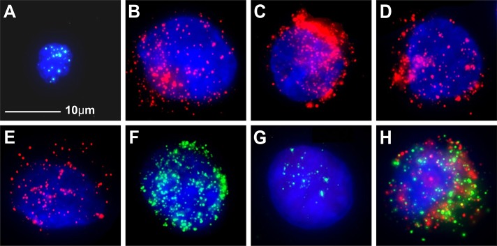
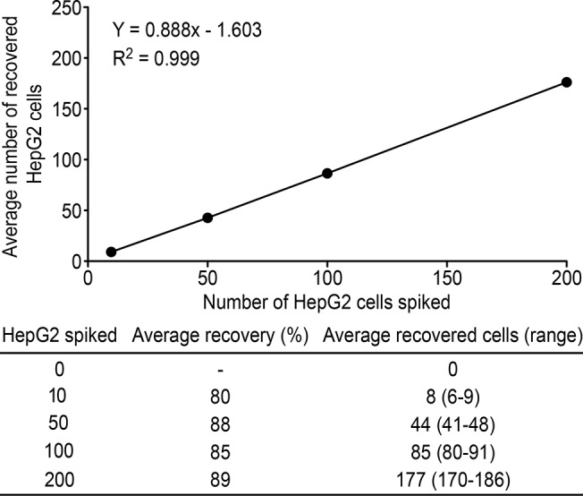
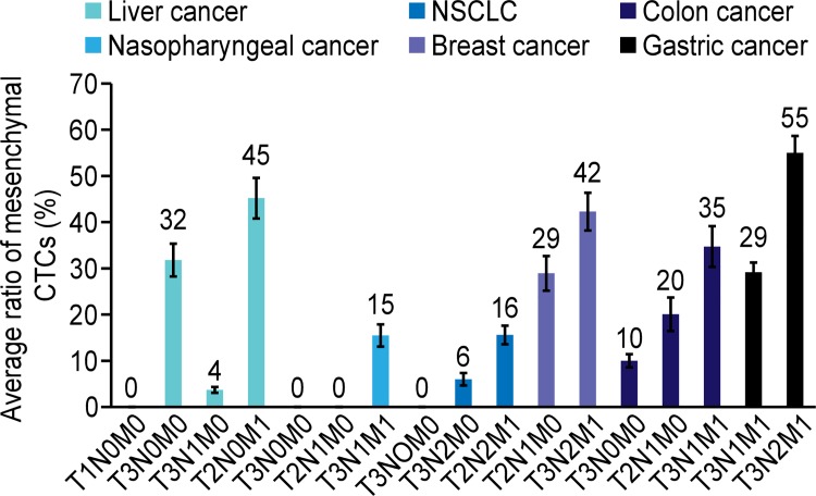
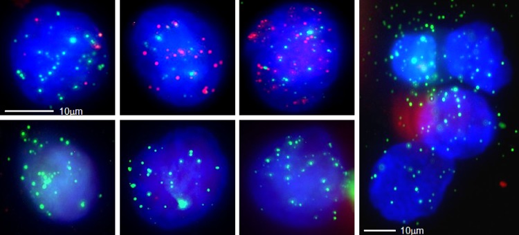
In cancer, epithelial-mesenchymal transition (EMT) is associated with metastasis. Characterizing EMT phenotypes in circulating tumor cells (CTCs) has been challenging because epithelial marker-based methods have typically been used for the isolation and detection of CTCs from blood samples. The aim of this study was to use the optimized CanPatrol CTC enrichment technique to classify CTCs using EMT markers in different types of cancers. The first step of this technique was to isolate CTCs via a filter-based method; then, an RNA in situ hybridization (RNA-ISH) method based on the branched DNA signal amplification technology was used to classify the CTCs according to EMT markers. Our results indicated that the efficiency of tumor cell recovery with this technique was at least 80%. When compared with the non-optimized method, the new method was more sensitive and more CTCs were detected in the 5-ml blood samples. To further validate the new method, 164 blood samples from patients with liver, nasopharyngeal, breast, colon, gastric cancer, or non-small-cell lung cancer (NSCLC) were collected for CTC isolation and characterization. CTCs were detected in 107 (65%) of 164 blood samples, and three CTC subpopulations were identified using EMT markers, including epithelial CTCs, biophenotypic epithelial/mesenchymal CTCs, and mesenchymal CTCs. Compared with the earlier stages of cancer, mesenchymal CTCs were more commonly found in patients in the metastatic stages of the disease in different types of cancers. Circulating tumor microemboli (CTM) with a mesenchymal phenotype were also detected in the metastatic stages of cancer. Classifying CTCs by EMT markers helps to identify the more aggressive CTC subpopulation and provides useful evidence for determining an appropriate clinical approach. This method is suitable for a broad range of carcinomas.
Conflict of interest statement
Figures
Similar articles
-
An improved strategy to detect the epithelial-mesenchymal transition process in circulating tumor cells in hepatocellular carcinoma patients.Hepatol Int. 2016 Jul;10(4):640-6. doi: 10.1007/s12072-016-9732-7. Epub 2016 Apr 26. Hepatol Int. 2016. PMID: 27115761
-
Analysis of the hormone receptor status of circulating tumor cell subpopulations based on epithelial-mesenchymal transition: a proof-of-principle study on the heterogeneity of circulating tumor cells.Oncotarget. 2016 Oct 4;7(40):65993-66002. doi: 10.18632/oncotarget.11787. Oncotarget. 2016. PMID: 27602758 Free PMC article.
-
Expression and clinical relevance of epithelial and mesenchymal markers in circulating tumor cells from colorectal cancer.Oncotarget. 2017 Feb 7;8(6):9293-9302. doi: 10.18632/oncotarget.14065. Oncotarget. 2017. PMID: 28030836 Free PMC article.
-
Circulating Tumor Cells and Implications of the Epithelial-to-Mesenchymal Transition.Adv Clin Chem. 2018;83:121-181. doi: 10.1016/bs.acc.2017.10.004. Epub 2017 Dec 21. Adv Clin Chem. 2018. PMID: 29304900 Review.
-
Expression of Epithelial Mesenchymal Transition and Cancer Stem Cell Markers in Circulating Tumor Cells.Adv Exp Med Biol. 2017;994:205-228. doi: 10.1007/978-3-319-55947-6_11. Adv Exp Med Biol. 2017. PMID: 28560676 Review.
Cited by
-
Platelets, immune cells and the coagulation cascade; friend or foe of the circulating tumour cell?Mol Cancer. 2021 Mar 31;20(1):59. doi: 10.1186/s12943-021-01347-1. Mol Cancer. 2021. PMID: 33789677 Free PMC article. Review.
-
Clinical significance of Ki67 and circulating tumor cells with an epithelial-mesenchymal transition phenotype in non-small cell lung cancer.Am J Transl Res. 2020 Jun 15;12(6):2916-2928. eCollection 2020. Am J Transl Res. 2020. PMID: 32655819 Free PMC article.
-
Clinical verification of vimentin/EpCAM immunolipid magnetic sorting system in monitoring CTCs in arterial and venous blood of advanced tumor.J Nanobiotechnology. 2021 Jun 16;19(1):185. doi: 10.1186/s12951-021-00929-x. J Nanobiotechnology. 2021. PMID: 34134721 Free PMC article.
-
Growth signaling autonomy in circulating tumor cells aids metastatic seeding.PNAS Nexus. 2024 Jan 25;3(2):pgae014. doi: 10.1093/pnasnexus/pgae014. eCollection 2024 Feb. PNAS Nexus. 2024. PMID: 38312224 Free PMC article.
-
Enumeration And Characterization Of Circulating Tumor Cells And Its Application In Advanced Gastric Cancer.Onco Targets Ther. 2019 Sep 25;12:7887-7896. doi: 10.2147/OTT.S223222. eCollection 2019. Onco Targets Ther. 2019. PMID: 31576146 Free PMC article.
References
-
- Fidler IJ. The pathogenesis of cancer metastasis: the seed and soil hypothesis revisited. Nat Rev Cancer. 2003; 3: 453–458. - PubMed
-
- Pantel K, Brakenhoff RH. Dissecting the metastatic cascade. Nat Rev Cancer. 2004; 4: 448–456. - PubMed
-
- Guarino M. Epithelial-mesenchymal transition and tumour invasion. Int J Biochem Cell Biol. 2007; 39: 2153–2160. - PubMed
Publication types
MeSH terms
Substances
LinkOut - more resources
Full Text Sources
Other Literature Sources
