. 2015 Dec;53(6):793-801.
doi: 10.1165/rcmb.2014-0422OC.

Efficacy of Leukadherin-1 in the Prevention of Hyperoxia-Induced Lung Injury in Neonatal Rats

Affiliations

Efficacy of Leukadherin-1 in the Prevention of Hyperoxia-Induced Lung Injury in Neonatal Rats

Jawahar Jagarapu et al. Am J Respir Cell Mol Biol. 2015 Dec.

Abstract

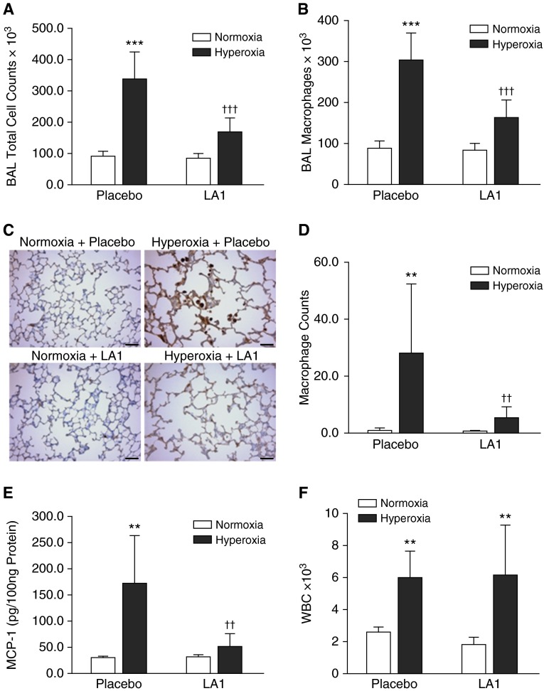
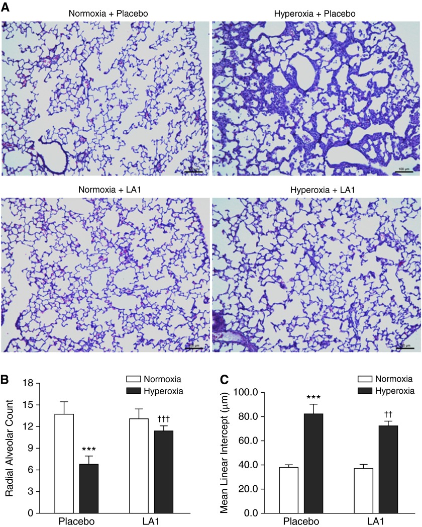
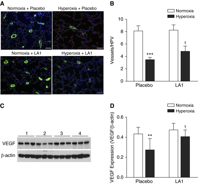
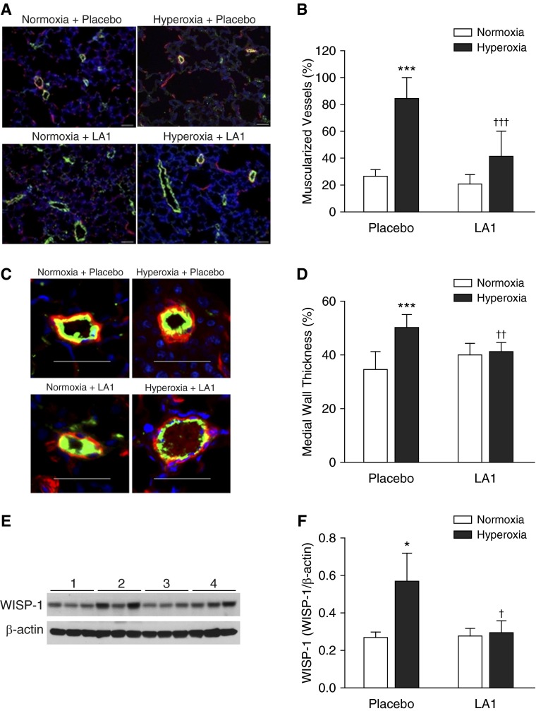
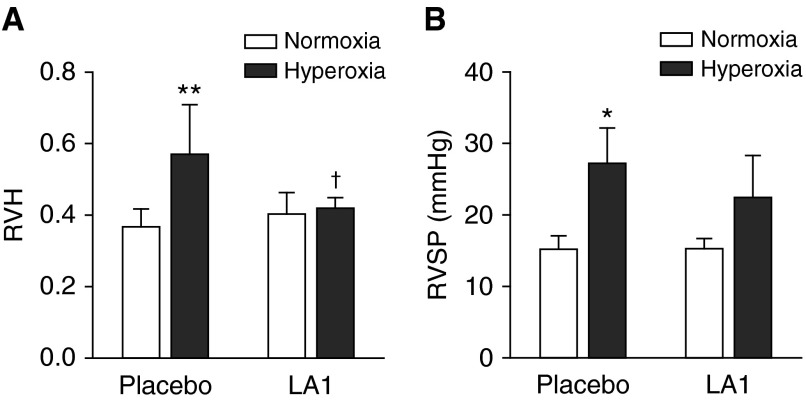
Lung inflammation plays a key role in the pathogenesis of bronchopulmonary dysplasia (BPD), a chronic lung disease of premature infants. The challenge in BPD management is the lack of effective and safe antiinflammatory agents. Leukadherin-1 (LA1) is a novel agonist of the leukocyte surface integrin CD11b/CD18 that enhances leukocyte adhesion to ligands and vascular endothelium and thus reduces leukocyte transendothelial migration and influx to the injury sites. Its functional significance in preventing hyperoxia-induced neonatal lung injury is unknown. We tested the hypothesis that administration of LA1 is beneficial in preventing hyperoxia-induced neonatal lung injury, an experimental model of BPD. Newborn rats were exposed to normoxia (21% O2) or hyperoxia (85% O2) and received twice-daily intraperitoneal injection of LA1 or placebo for 14 days. Hyperoxia exposure in the presence of the placebo resulted in a drastic increase in the influx of neutrophils and macrophages into the alveolar airspaces. This increased leukocyte influx was accompanied by decreased alveolarization and angiogenesis and increased pulmonary vascular remodeling and pulmonary hypertension (PH), the pathological hallmarks of BPD. However, administration of LA1 decreased macrophage infiltration in the lungs during hyperoxia. Furthermore, treatment with LA1 improved alveolarization and angiogenesis and decreased pulmonary vascular remodeling and PH. These data indicate that leukocyte recruitment plays an important role in the experimental model of BPD induced by hyperoxia. Targeting leukocyte trafficking using LA1, an integrin agonist, is beneficial in preventing lung inflammation and protecting alveolar and vascular structures during hyperoxia. Thus, targeting integrin-mediated leukocyte recruitment and inflammation may provide a novel strategy in preventing and treating BPD in preterm infants.

Keywords: BPD; LA1; hyperoxia; inflammation; integrin.


Figures

Figure 1.
Figure 1.
Administration of leukadherin-1 (LA1) decreases leukocyte influx into the lung and monocyte chemoattractant protein-1 (MCP-1) expression. Total cell counts (A) and macrophage counts (B) in bronchoalveolar lavage (BAL) were increased in hyperoxia- and placebo-exposed animals. However, these were significantly decreased by treatment with LA1. Immunostaining for Mac3 was performed on lung tissue sections (C), and the average numbers of macrophages in alveolar airspaces were determined from 10 random images taken under the high-power view on each lung section (D). There was an increased macrophage count in the placebo-treated hyperoxia group, but this number was drastically decreased upon treatment with LA1. (E) MCP-1 in lung homogenates was increased in the hyperoxia + placebo group, but it was decreased by treatment with LA1. (F) White blood cells (WBCs) were counted in peripheral blood. Hyperoxia exposure significantly increased WBC count in both the placebo and LA1-treated groups as compared with normoxic groups. Scale bar: 50 μm (n = 4 to 5 per group). **P < 0.01 and ***P < 0.001 compared with normoxic groups; ††P < 0.01 and †††P < 0.001 compared with the hyperoxia + placebo group.
Figure 2.
Figure 2.
Treatment with LA1 improves alveolar development. (A) On histological examination, lungs from placebo-treated and hyperoxia-exposed rats displayed an impairment of alveolarization with fewer, larger, and simplified alveoli. Treatment with LA1 improved alveolar structures that appeared similar to the normoxic lungs. On morphometric assessment, placebo-treated animals exposed to hyperoxia showed a decrease in radial alveolar count (RAC) (B) and increased mean linear intercept (MLI) (C) as compared with normoxic animals. Administration of LA1 during hyperoxia significantly increased RAC and decreased MLI. Scale bar: 100 μm (n = 5 per group). ***P < 0.001 compared with normoxic groups; ††P < 0.01 and †††P < 0.001 compared with the hyperoxia + placebo group.
Figure 3.
Figure 3.
LA1 administration improves vascular development. (A) Double immunofluorescence staining with an anti–von Willebrand factor (vWF) antibody (green signals), anti–α-smooth muscle actin (α-SMA) antibody (red signal), and 4′,6-diamidino-2-phenylindole (DAPI) nuclear staining (blue signals) was performed on lung tissue sections. (B) Vascular density was determined by counting vWF-positive vessels (<50 μm) on 10 random images from each lung section. The vascular density was significantly decreased in hyperoxia + placebo lungs compared with normoxia lungs. Treatment with LA1 modestly increased vascular density in hyperoxia-exposed animals. (C) Representative photo images of Western blots for vascular endothelial growth factor (VEGF) and β-actin expression. (D) Densitometry analysis demonstrated that VEGF expression was significantly decreased in hyperoxia + placebo lungs but was increased by treatment with LA1. Scale bar: 50 μm. **P < 0.01, and ***P < 0.001 compared with normoxic groups (n = 5 per group). P < 0.05 compared with hyperoxia plus placebo group. HPV, high-power view.
Figure 4.
Figure 4.
Treatment with LA1 attenuates hyperoxia-induced pulmonary vascular remodeling. (A) Double-immunofluorescence staining for vWF (green signals) and α-SMA (red signals) and DAPI nuclear staining (blue signals). (B) The percentage of muscularized peripheral pulmonary vessels (≥50% of circumference) was significantly increased in lungs from the hyperoxia + placebo group. Administration of LA1 significantly decreased vascular muscularization in hyperoxia-exposed animals. (C) Medial wall thickness (MWT) was assessed on 20 peripheral vessels (<50 μm in diameter) on each lung section. (D) Hyperoxia significantly increased MWT in lungs of placebo-treated animals. However, treatment with LA1 significantly decreased MWT during hyperoxia. (E) Representative photo images of Western blots for Wnt1-inducible signaling protein-1 (WISP-1) and β-actin expression. (F) Densitometry analysis demonstrated that WISP-1 expression was significantly increased in the hyperoxia + placebo lungs and was decreased by administration of LA1. Scale bar: 50 μm. *P < 0.05 and ***P < 0.001 compared with normoxic groups; P < 0.05, ††P < 0.01, and †††P < 0.001 compared with hyperoxia + placebo group (n = 5 per group).
Figure 5.
Figure 5.
Effects of LA1 on hyperoxia-induced pulmonary hypertension (PH). Right ventricular hypertrophy (RVH) (A) and right ventricular systolic pressure (RVSP) (B) were assessed as indices of PH. Hyperoxia exposure resulted in RVH in placebo-treated rats. Administration of LA1 significantly decreased RVH during hyperoxia. Hyperoxia also increased RVSP in placebo-exposed rats. Treatment with LA1 resulted in a slightly decreased RVSP during hyperoxia, but this did not reach statistical significance. *P < 0.05 and **P < 0.01 compared with normoxic groups; P < 0.05 compared with hyperoxia + placebo group (n = 5 per group).

Similar articles

Cited by

References

    1. Jobe AH, Bancalari E. Bronchopulmonary dysplasia. Am J Respir Crit Care Med. 2001;163:1723–1729. - PubMed
    1. Bhandari A, Bhandari V. Pitfalls, problems, and progress in bronchopulmonary dysplasia. Pediatrics. 2009;123:1562–1573. - PubMed
    1. Thebaud B, Abmen SH. Bronchpulmonary dysplasia: where have all the vessels gone? Roles of angiogenic growth factors in chronic lung disease. Am J Respir Crit Care Med. 2007;175:978–985. - PMC - PubMed
    1. Stenmark KR, Abman SH. Lung vascular development: implications for pathogenesis of bronchopulmonary dysplasia. Annu Rev Physiol. 2005;67:623–661. - PubMed
    1. Speer CP. Inflammation and bronchopulmonary dysplasia: a continuing story. Semin Fetal Neonatal Med. 2006;11:354–362. - PubMed

Publication types

MeSH terms