Review
. 2015 Jun:32:171-80.
doi: 10.1016/j.gde.2015.03.004. Epub 2015 Apr 21.

Islet formation in mice and men: lessons for the generation of functional insulin-producing β-cells from human pluripotent stem cells

Affiliations
Review

Islet formation in mice and men: lessons for the generation of functional insulin-producing β-cells from human pluripotent stem cells

Gopika Nair et al. Curr Opin Genet Dev. 2015 Jun.

Abstract

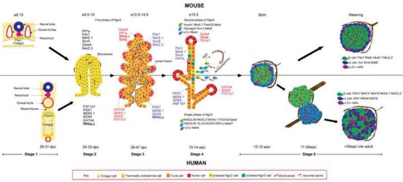
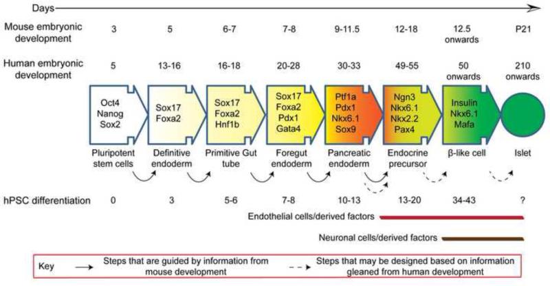
The Islets of Langerhans are crucial 'micro-organs' embedded in the glandular exocrine pancreas that regulate nutrient metabolism. They not only synthesize, but also secrete endocrine hormones in a modulated fashion in response to physiologic metabolic demand. These highly sophisticated structures with intricate organization of multiple cell types, namely endocrine, vascular, neuronal and mesenchymal cells, have evolved to perform this task to perfection over time. Not surprisingly, islet architecture and function are dissimilar between humans and typically studied model organisms, such as rodents and zebrafish. Further, recent findings also suggest noteworthy differences in human islet development from that in mouse, including delayed appearance and gradual resolution of key differentiation markers, a single-phase of endocrine differentiation, and prenatal association of developing islets with neurovascular milieu. In light of these findings, it is imperative that a systematic study is undertaken to compare islet development between human and mouse. Illuminating inter-species differences in islet development will likely be critical in furthering our pursuit to generate an unlimited supply of truly functional and fully mature β-cells from human pluripotent stem cell (hPSC) sources for therapeutic purposes.


Figures

Fig. 1
Fig. 1
Illustration of pancreas morphogenesis in mouse and human. The top half of the figure depicts mouse while the bottom half portrays human development. Stage 1: Pancreas specification: appearance of Pdx1 marks the presumptive pancreatic region in the foregut epithelium. In mouse Pdx1 appears before gut closure when the notochord is in contact with the gut, but in human, PDX1 is delayed and appears only after separation of notochord and aorta from the gut. Stage 2: Formation of pancreatic epithelium: Following Pdx1 expression and Ptf1a activation, pancreatic buds containing MPCs grow into the surrounding mesenchyme. MPCs exhibit similar transcript profile between mouse and human with the exception of NKX2.2. Microlumens are already visible at this stage. Stage 3: Segregation of the tip and trunk domain: MPCs differentiate into pro-acinar tip cells (indicated by light red) and bipotential epithelial cords(light orange) due to opposing functions of Ptf1a and Nkx6.1. This process is dynamic with rampant tubulogenesis. In human, the separation occurs gradually with the tip cells still expressing NKX6.1 and SOX9 at the corresponding age in mice. Stage 4: Endocrine differentiation (also secondary transition in mouse). Terminal differentiation of tips to acinar fate and trunk cells to endocrine/duct fate occur during this period. The second wave of Ngn3 expression coincides with this period in the mouse, whereas in human, this period occurs after embryogenesis and a single phase of Ngn3 expression is observed. The time point indicated denote the peak period of endocrine differentiation in both species. Stage 5: Islet morphogenesis. In mouse, sympathetic axons regulate islet cell clustering by β-adrenergic signaling while a parallel is unknown in human. Moreover, islet formation along with vascularization is completed only at birth in the mouse, whereas in human, islet morphogenesis extends through the second and third trimester but is completed before birth. Initially fetal human islets resemble mouse islets in its architecture. Then, they opens up to give rise to juxtaposed homotypic α/β clusters, and finally acquire the intermingled architecture of adult islets in the last trimester. Innervation of mouse islets is completed at weaning (orange arrow), whereas human islets lose their dense pre-natal neuronal associations. In addition, axons contact vasculature in human adult islets (orange arrowhead). The approximate embryonic/fetal age at which the various stages occur are designated respectively for the two species. Progressively darker shades of the colors representing epithelial and endocrine cells indicate differentiation to more committed fates over time. e-embryonic day; dpc-days post conception; wpc-weeks post conception
Fig. 2
Fig. 2
Timeline of pancreas development, in vivo, in mouse and human compared with in vitro differentiation of β-cells from hPSCs. hPSC differentiation protocols have been developed largely based on information gained from pancreas development in mice. Current protocols generate sub-par β-like cells that have modest glucose responsiveness but do not resemble mature β-cells of an islet in their metabolic and secretory properties. New data from human pancreas development, such as involvement of neuronal and endothelial cells in later stages of organogenesis when β-cell maturation predominantly occurs, may be used to instruct better differentiation protocols that result in bona fide β-cells. The timeline of hPSC differentiation is based on two recent studies; Pagliuca et al. [66]and Rezania et al.[67]

References

    1. Kushner Jake A, MacDonald Patrick E, Atkinson Mark A. Stem Cells to Insulin Secreting Cells: Two Steps Forward and Now a Time to Pause? Cell Stem Cell. 2014;15(5):535–536. [The review discusses the shortcomings in two recent studies that report sucessful generation of insulin secreting cells invitro from human pluripotent stem cells.] - PubMed
    1. Pagliuca FW, Melton DA. How to make a functional β-cell. Development. 2013;140(12):2472–2483. - PMC - PubMed
    1. Hay CW, Docherty K. Comparative Analysis of Insulin Gene Promoters: Implications for Diabetes Research. Diabetes. 2006;55(12):3201–3213. - PubMed
    1. McCulloch LJ, van de Bunt M, Braun M, et al. GLUT2 (SLC2A2) is not the principal glucose transporter in human pancreatic beta cells: Implications for understanding genetic association signals at this locus. Molecular Genetics and Metabolism. 2011;104(4):648–653. - PubMed
    1. De Vos A, Heimberg H, Quartier E, et al. Human and rat beta cells differ in glucose transporter but not in glucokinase gene expression. The Journal of Clinical Investigation. 1995;96(5):2489–2495. - PMC - PubMed

Publication types