. 2015 Apr 24;10(4):e0124922.
doi: 10.1371/journal.pone.0124922. eCollection 2015.

View-Angle Tilting and Slice-Encoding Metal Artifact Correction for Artifact Reduction in MRI: Experimental Sequence Optimization for Orthopaedic Tumor Endoprostheses and Clinical Application

Affiliations

View-Angle Tilting and Slice-Encoding Metal Artifact Correction for Artifact Reduction in MRI: Experimental Sequence Optimization for Orthopaedic Tumor Endoprostheses and Clinical Application

Pia M Jungmann et al. PLoS One. .

Abstract

Background: MRI plays a major role in follow-up of patients with malignant bone tumors. However, after limb salvage surgery, orthopaedic tumor endoprostheses might cause significant metal-induced susceptibility artifacts.

Purposes: To evaluate the benefit of view-angle tilting (VAT) and slice-encoding metal artifact correction (SEMAC) for MRI of large-sized orthopaedic tumor endoprostheses in an experimental model and to demonstrate clinical benefits for assessment of periprosthetic soft tissue abnormalities.

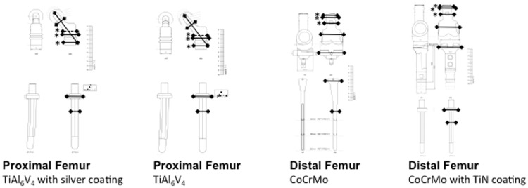
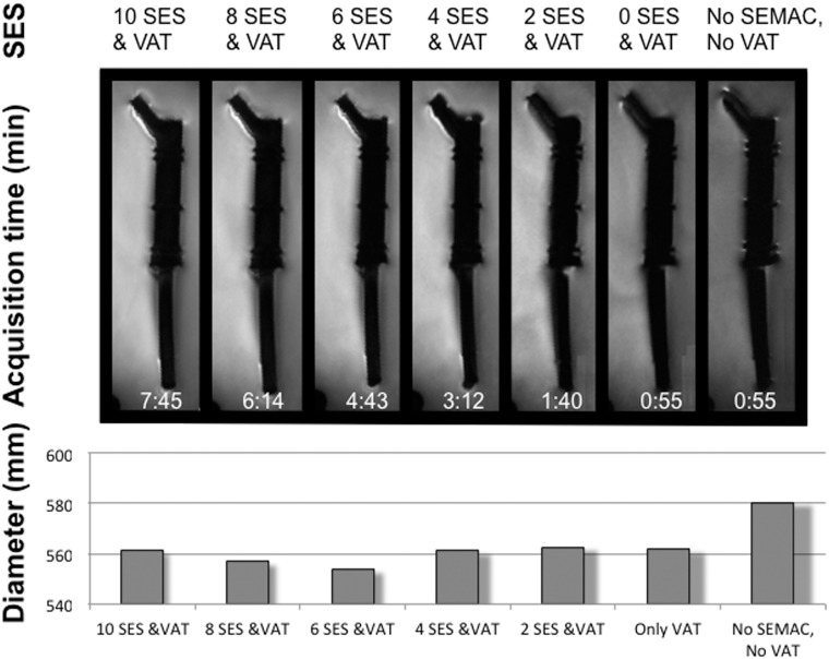
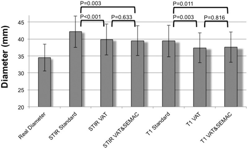
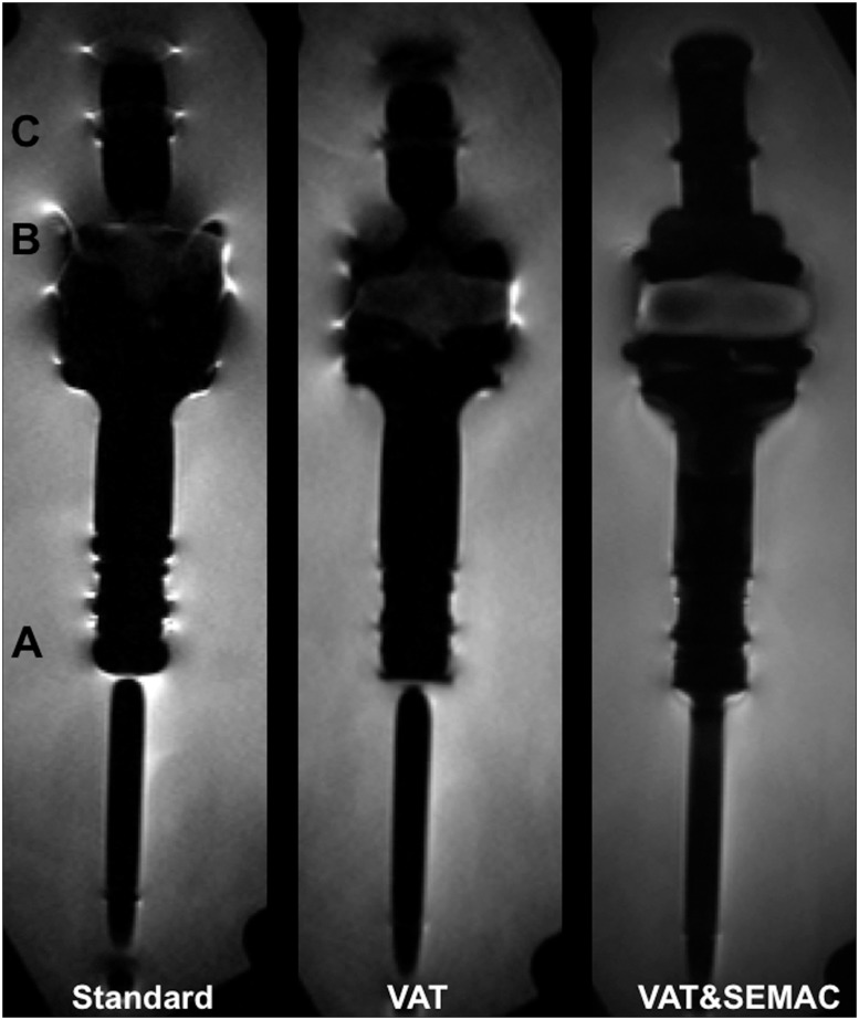
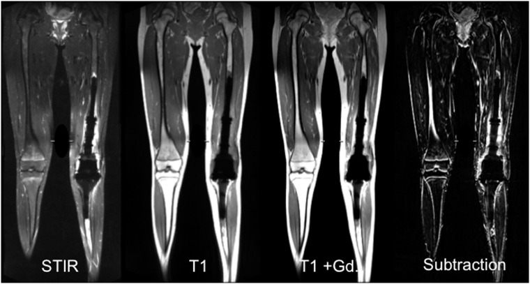
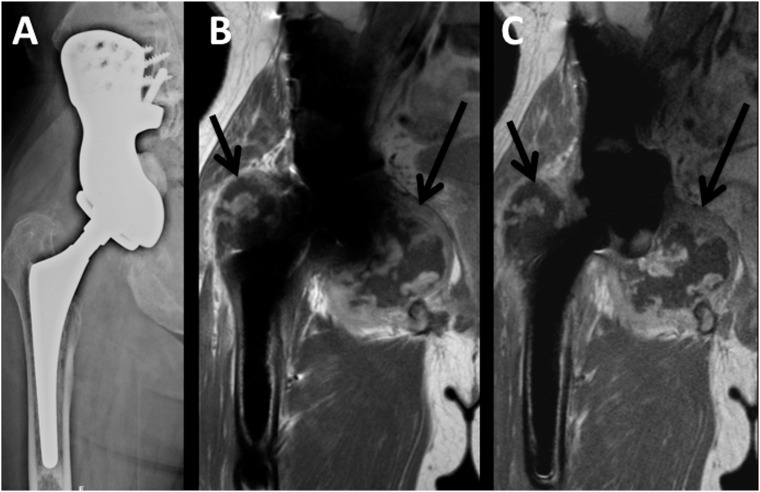
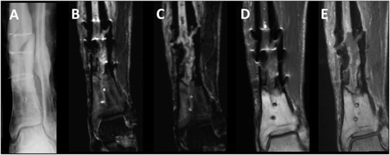
Methods: In an experimental setting, tumor endoprostheses (n=4) were scanned at 1.5T with three versions of optimized high-bandwidth turbo-spin-echo pulse sequences: (i) standard, (ii) VAT and (iii) combined VAT and SEMAC (VAT&SEMAC). Pulse sequences included coronal short-tau-inversion-recovery (STIR), coronal T1-weighted (w), transverse T1-w and T2-w TSE sequences. For clinical evaluation, VAT&SEMAC was compared to conventional metal artifact-reducing MR sequences (conventional MR) in n=25 patients with metal implants and clinical suspicion of tumor recurrence or infection. Diameters of artifacts were measured quantitatively. Qualitative parameters were assessed on a five-point scale (1=best, 5=worst): "image distortion", "artificial signal changes at the edges" and "diagnostic confidence". Imaging findings were correlated with pathology. T-tests and Wilcoxon-signed rank tests were used for statistical analyses.

Results: The true size of the prostheses was overestimated on MRI (P<0.05). A significant reduction of artifacts was achieved by VAT (P<0.001) and VAT&SEMAC (P=0.003) compared to the standard group. Quantitative scores improved in the VAT and VAT&SEMAC group (P<0.05). On clinical MR images, artifact diameters were significantly reduced in the VAT&SEMAC-group as compared with the conventional-group (P<0.001). Distortion and artificial signal changes were reduced and diagnostic confidence improved (P<0.05). In two cases, tumor-recurrence, in ten cases infection and in thirteen cases other pathologies were diagnosed.

Conclusions: Significant reduction of metallic artifacts was achieved by VAT and SEMAC. Clinical results suggest, that these new techniques will be beneficial for detecting periprosthetic pathologies during postoperative follow-up.


Conflict of interest statement

Competing Interests: The co-author M.N. is an employee of Siemens and was involved in the technical development of the metal-artifact reducing pulse sequences. He did not contribute to, access or influence the design, performance or analysis of this study. This does not alter the authors' adherence to PLOS ONE policies on sharing data and materials.

Figures

Fig 1
Fig 1. Experimental model.
Tumor endoprostheses were embedded in water. Specifications and materials are indicated on the left. Central slices of coronal STIR and coronal T1-w MR pulse sequences are presented for each technique (standard; VAT, view-angle tilting; VAT&SEMAC, view-angle tilting and slice-encoding metal artifact correction).
Fig 2
Fig 2. True diameters of orthopaedic tumor endoprostheses.
True diameters were measured at six standardized spots as indicated and confirmed on original sketches (continuous lines). Asterisks indicate three standardized spots that were used for comparisons of different implant materials; broken lines indicate diameters that were additionally measured for this purpose.
Fig 3
Fig 3. Reduction of slice-encoding steps (SES) for slice-encoding metal artifact correction (SEMAC).
SES were reduced from 10 to 0 and assessed quantitatively and qualitatively. No SEMAC is equivalent to 0 SES. Acquisition time for all SES values is shown in the images. For qualitative evaluation, artifact diameters (mm) were measured on all MR images. VAT, view-angle tilting. SEMAC; slice-encoding metal artifact correction.
Fig 4
Fig 4. Quantitative results for coronal MR pulse sequences (STIR and T1-w sequences).
Diameter measurements were performed at six standardized levels on all MR images and compared to true diameters of the tumor endoprostheses. Study groups were a standard group (Standard) without application of VAT (view-angle tilting) or SEMAC (slice-encoding metal artifact correction) but otherwise identical pulse sequences, a group where VAT was applied (VAT; no SEMAC) and a group where both techniques, VAT and SEMAC, were applied (VAT&SEMAC). Mean values ±SEM are presented. True diameters were significantly smaller than artifact diameters in all groups (P<0.05).
Fig 5
Fig 5. Coronal STIR images of one tumor endoprosthesis are presented for all groups (standard group; VAT (view-angle tilting) group; VAT&SEMAC (view-angle tilting and slice-encoding metal artifact correction) group).
Exemplarily, “A” indicates the interface between the collar of the stem and the stem of the prosthesis where the qualitative parameter “geometric distortion” was evaluated. “B” indicates the true position of the polyethylene component, which is interposed between the distal femoral component and the proximal tibial component of the endoprosthesis. “C” indicates the signal loss and signal pile-up artifacts at the edges of the prosthesis.
Fig 6
Fig 6. Example of the large size of the field of view.
A Large field of view is needed for postoperative follow-up MR imaging in tumor patients. MR imaging was performed in a young, female patient (15 years old) after resection of an osteosarcoma at the left distal femur and implantation of a combined femoral and tibial replacement. Pulse sequences presented include a coronal STIR sequence and a coronal T1-w sequence without and with contrast application and a subtraction image, all using view-angle tilting and slice-encoding metal artifact correction with 6 slice encoding steps.
Fig 7
Fig 7. Tumor recurrence.
Hip endoprosthesis with tumor recurrence of a chondrosarcoma in a 45 year old male patient. (A) a.p. radiograph; (B) Coronal contrast enhanced T1-w image with conventional artifact reducing MR techniques; (C) Coronal contrast enhanced T1-w image with VAT&SEMAC techniques.
Fig 8
Fig 8. Infection.
Posttraumatic pseudarthrosis at the left distal tibia with infection and intramedullary abscess formation in a 27 year old male patient with remaining cerclage fixation. (A) a.p. radiograph; (B) conventional coronal STIR image; (C) VAT&SEMAC coronal STIR image; (D) conventional coronal contrast enhanced T1-w image; (E) VAT&SEMAC contrast enhanced coronal T1-w image.

References

    1. Choong PF, Sim FH, Pritchard DJ, Rock MG, Chao EY. Megaprostheses after resection of distal femoral tumors. A rotating hinge design in 30 patients followed for 2–7 years. Acta Orthop Scand. 1996;67(4):345–51. - PubMed
    1. Eckardt JJ, Eilber FR, Rosen G, Mirra JM, Dorey FJ, Ward WG, et al. Endoprosthetic replacement for stage IIB osteosarcoma. Clin Orthop Relat Res. 1991(270):202–13. - PubMed
    1. Kawai A, Muschler GF, Lane JM, Otis JC, Healey JH. Prosthetic knee replacement after resection of a malignant tumor of the distal part of the femur. Medium to long-term results. J Bone Joint Surg Am. 1998;80(5):636–47. - PubMed
    1. Morris HG, Capanna R, Campanacci D, Del Ben M, Gasbarrini A. Modular endoprosthetic replacement after total resection of the femur for malignant tumour. Int Orthop. 1994;18(2):90–5. - PubMed
    1. Sim FH, Frassica FJ, Chao EY. Orthopaedic management using new devices and prostheses. Clin Orthop Relat Res. 1995(312):160–72. - PubMed

Publication types