Chloroform-Methanol Residue of Coxiella burnetii Markedly Potentiated the Specific Immunoprotection Elicited by a Recombinant Protein Fragment rOmpB-4 Derived from Outer Membrane Protein B of Rickettsia rickettsii in C3H/HeN Mice
- PMID: 25909586
- PMCID: PMC4409375
- DOI: 10.1371/journal.pone.0124664
Chloroform-Methanol Residue of Coxiella burnetii Markedly Potentiated the Specific Immunoprotection Elicited by a Recombinant Protein Fragment rOmpB-4 Derived from Outer Membrane Protein B of Rickettsia rickettsii in C3H/HeN Mice
Abstract
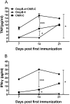
The obligate intracellular bacteria, Rickettsia rickettsii and Coxiella burnetii, are the potential agents of bio-warfare/bio-terrorism. Here C3H/HeN mice were immunized with a recombinant protein fragment rOmp-4 derived from outer membrane protein B, a major protective antigen of R. rickettsii, combined with chloroform-methanol residue (CMR) extracted from phase I C. burnetii organisms, a safer Q fever vaccine. These immunized mice had significantly higher levels of IgG1 and IgG2a to rOmpB-4 and interferon-γ (IFN-γ) and tumor necrosis factor-α (TNF-α), two crucial cytokines in resisting intracellular bacterial infection, as well as significantly lower rickettsial loads and slighter pathological lesions in organs after challenge with R. rickettsii, compared with mice immunized with rOmpB-4 or CMR alone. Additionally, after challenge with C. burnetii, the coxiella loads in the organs of these mice were significantly lower than those of mice immunized with rOmpB-4 alone. Our results prove that CMR could markedly potentiate enhance the rOmpB-4-specific immunoprotection by promoting specific and non-specific immunoresponses and the immunization with the protective antigen of R. rickettsii combined with CMR of C. burnetii could confer effective protection against infection of R. rickettsii or C. burnetii.
Conflict of interest statement
Figures


Similar articles
-
Enhanced protection against Rickettsia rickettsii infection in C3H/HeN mice by immunization with a combination of a recombinant adhesin rAdr2 and a protein fragment rOmpB-4 derived from outer membrane protein B.Vaccine. 2015 Feb 18;33(8):985-92. doi: 10.1016/j.vaccine.2015.01.017. Epub 2015 Jan 15. Vaccine. 2015. PMID: 25597943
-
Surface protein Adr2 of Rickettsia rickettsii induced protective immunity against Rocky Mountain spotted fever in C3H/HeN mice.Vaccine. 2014 Apr 11;32(18):2027-33. doi: 10.1016/j.vaccine.2014.02.057. Epub 2014 Feb 28. Vaccine. 2014. PMID: 24582636
-
Enhanced protection against Q fever in BALB/c mice elicited by immunization of chloroform-methanol residue of Coxiella burnetii via intratracheal inoculation.Vaccine. 2019 Sep 24;37(41):6076-6084. doi: 10.1016/j.vaccine.2019.08.041. Epub 2019 Aug 30. Vaccine. 2019. PMID: 31477436
-
Vaccines against coxiellosis and Q fever. Development of a chloroform:methanol residue subunit of phase I Coxiella burnetti for the immunization of animals.Ann N Y Acad Sci. 1992 Jun 16;653:88-111. doi: 10.1111/j.1749-6632.1992.tb19633.x. Ann N Y Acad Sci. 1992. PMID: 1626897 Review.
-
Adaptive immunity to the obligate intracellular pathogen Coxiella burnetii.Immunol Res. 2009;43(1-3):138-48. doi: 10.1007/s12026-008-8059-4. Immunol Res. 2009. PMID: 18813881 Free PMC article. Review.
Cited by
-
Effects of Mycobacterium vaccae vaccine in a mouse model of tuberculosis: protective action and differentially expressed genes.Mil Med Res. 2020 Jun 3;7(1):25. doi: 10.1186/s40779-020-00258-4. Mil Med Res. 2020. PMID: 32493477 Free PMC article.
-
Animal Models of Tuberculosis Vaccine Research: An Important Component in the Fight against Tuberculosis.Biomed Res Int. 2020 Jan 2;2020:4263079. doi: 10.1155/2020/4263079. eCollection 2020. Biomed Res Int. 2020. PMID: 32025519 Free PMC article. Review.
-
Amblyomma sculptum Salivary PGE2 Modulates the Dendritic Cell-Rickettsia rickettsii Interactions in vitro and in vivo.Front Immunol. 2019 Feb 4;10:118. doi: 10.3389/fimmu.2019.00118. eCollection 2019. Front Immunol. 2019. PMID: 30778355 Free PMC article.
-
Diagnostic accuracy of Mycobacterium tuberculosis-specific triple-color FluoroSpot assay in differentiating tuberculosis infection status in febrile patients with suspected tuberculosis.Front Immunol. 2025 Jan 8;15:1462222. doi: 10.3389/fimmu.2024.1462222. eCollection 2024. Front Immunol. 2025. PMID: 39845975 Free PMC article.
-
Soluble antigens derived from Coxiella burnetii elicit protective immunity in three animal models without inducing hypersensitivity.Cell Rep Med. 2021 Dec 6;2(12):100461. doi: 10.1016/j.xcrm.2021.100461. eCollection 2021 Dec 21. Cell Rep Med. 2021. PMID: 35028605 Free PMC article.
References
-
- Walker DH, Crawford CG, Cain BG. Rickettsial infection of the pulmonary microcirculation: the basis for interstitial pneumonitis in Rocky Mountain spotted fever. Human Pathology. 1980;11(3):263–72. . - PubMed
-
- Horney LF, Walker DH. Meningoencephalitis as a major manifestation of Rocky Mountain spotted fever. Southern Medical Journal. 1988;81(7):915–8. . - PubMed
-
- Harrell GT, Aikawa JK. Pathogenesis of circulatory failure in Rocky Mountain spotted fever: alterations in the blood volume and the thiocyanate space at various stages of the disease. Archives of Internal Medicine. 1949;83(3):331 . - PubMed
-
- Silverman DJ. Adherence of platelets to human endothelial cells infected by Rickettsia rickettsii . Journal of Infectious Diseases. 1986;153(4):694–700. . - PubMed
Publication types
MeSH terms
Substances
LinkOut - more resources
Full Text Sources
Other Literature Sources

