

Characterization of Burkholderia pseudomallei Strains Using a Murine Intraperitoneal Infection Model and In Vitro Macrophage Assays
- PMID: 25909629
- PMCID: PMC4409376
- DOI: 10.1371/journal.pone.0124667
Characterization of Burkholderia pseudomallei Strains Using a Murine Intraperitoneal Infection Model and In Vitro Macrophage Assays
Abstract
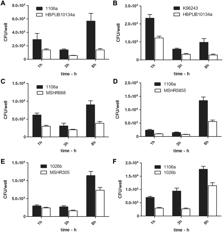
Burkholderia pseudomallei, the etiologic agent of melioidosis, is a gram-negative facultative intracellular bacterium. This bacterium is endemic in Southeast Asia and Northern Australia and can infect humans and animals by several routes. It has also been estimated to present a considerable risk as a potential biothreat agent. There are currently no effective vaccines for B. pseudomallei, and antibiotic treatment can be hampered by nonspecific symptomology, the high incidence of naturally occurring antibiotic resistant strains, and disease chronicity. Accordingly, there is a concerted effort to better characterize B. pseudomallei and its associated disease. Before novel vaccines and therapeutics can be tested in vivo, a well characterized animal model is essential. Previous work has indicated that mice may be a useful animal model. In order to develop standardized animal models of melioidosis, different strains of bacteria must be isolated, propagated, and characterized. Using a murine intraperitoneal (IP) infection model, we tested the virulence of 11 B. pseudomallei strains. The IP route offers a reproducible way to rank virulence that can be readily reproduced by other laboratories. This infection route is also useful in distinguishing significant differences in strain virulence that may be masked by the exquisite susceptibility associated with other routes of infection (e.g., inhalational). Additionally, there were several pathologic lesions observed in mice following IP infection. These included varisized abscesses in the spleen, liver, and haired skin. This model indicated that commonly used laboratory strains of B. pseudomallei (i.e., K96243 and 1026b) were significantly less virulent as compared to more recently acquired clinical isolates. Additionally, we characterized in vitro strain-associated differences in virulence for macrophages and described a potential inverse relationship between virulence in the IP mouse model of some strains and in the macrophage phagocytosis assay. Strains which were more virulent for mice (e.g., HBPU10304a) were often less virulent in the macrophage assays, as determined by several parameters such as intracellular bacterial replication and host cell cytotoxicity.
Conflict of interest statement
Figures
Similar articles
-
Characterization of pathogenesis of and immune response to Burkholderia pseudomallei K96243 using both inhalational and intraperitoneal infection models in BALB/c and C57BL/6 mice.PLoS One. 2017 Feb 24;12(2):e0172627. doi: 10.1371/journal.pone.0172627. eCollection 2017. PLoS One. 2017. PMID: 28235018 Free PMC article.
-
Comparative virulence of three different strains of Burkholderia pseudomallei in an aerosol non-human primate model.PLoS Negl Trop Dis. 2021 Feb 11;15(2):e0009125. doi: 10.1371/journal.pntd.0009125. eCollection 2021 Feb. PLoS Negl Trop Dis. 2021. PMID: 33571211 Free PMC article.
-
Comparison of the early host immune response to two widely diverse virulent strains of Burkholderia pseudomallei that cause acute or chronic infections in BALB/c mice.Microb Pathog. 2015 Sep;86:53-63. doi: 10.1016/j.micpath.2015.07.004. Epub 2015 Jul 7. Microb Pathog. 2015. PMID: 26162294
-
The molecular and cellular basis of pathogenesis in melioidosis: how does Burkholderia pseudomallei cause disease?FEMS Microbiol Rev. 2009 Nov;33(6):1079-99. doi: 10.1111/j.1574-6976.2009.00189.x. Epub 2009 Aug 5. FEMS Microbiol Rev. 2009. PMID: 19732156 Review.
-
An objective approach for Burkholderia pseudomallei strain selection as challenge material for medical countermeasures efficacy testing.Front Cell Infect Microbiol. 2012 Sep 26;2:120. doi: 10.3389/fcimb.2012.00120. eCollection 2012. Front Cell Infect Microbiol. 2012. PMID: 23057010 Free PMC article. Review.
Cited by
-
Distribution of Serological Response to Burkholderia pseudomallei in Swine from Three Provinces of Vietnam.Int J Environ Res Public Health. 2020 Jul 18;17(14):5203. doi: 10.3390/ijerph17145203. Int J Environ Res Public Health. 2020. PMID: 32708490 Free PMC article.
-
Efficacy of Treatment with the Antibiotic Novobiocin against Infection with Bacillus anthracis or Burkholderia pseudomallei.Antibiotics (Basel). 2022 Nov 23;11(12):1685. doi: 10.3390/antibiotics11121685. Antibiotics (Basel). 2022. PMID: 36551342 Free PMC article.
-
Characterization of the murine macrophage response to infection with virulent and avirulent Burkholderia species.BMC Microbiol. 2015 Nov 6;15:259. doi: 10.1186/s12866-015-0593-3. BMC Microbiol. 2015. PMID: 26545875 Free PMC article.
-
A Novel Virulent Clinical Isolate of Burkholderia pseudomallei Imported from Thailand Exhibiting Resistance to Trimethoprim-Sulfamethoxazole.Am J Trop Med Hyg. 2024 Oct 22;112(1):116-123. doi: 10.4269/ajtmh.24-0231. Print 2025 Jan 8. Am J Trop Med Hyg. 2024. PMID: 39437772
-
Proteomic Analysis of Non-human Primate Peripheral Blood Mononuclear Cells During Burkholderia mallei Infection Reveals a Role of Ezrin in Glanders Pathogenesis.Front Microbiol. 2021 Apr 22;12:625211. doi: 10.3389/fmicb.2021.625211. eCollection 2021. Front Microbiol. 2021. PMID: 33967974 Free PMC article.
References
-
- Vietri NJ, DeShazer D (2007) Melioidosis In: Dembek Z, editor. Textbook of Military Medicine: Medical Aspect of Biological Warfare. Washington, D.C.: Borden Institute Walter Reed Army Medical Center; pp. 147–166.
Publication types
MeSH terms
Substances
LinkOut - more resources
Full Text Sources
Other Literature Sources
