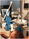
In Vivo Multiphoton Microscopy of Basal Cell Carcinoma
- PMID: 25909650
- PMCID: PMC4607557
- DOI: 10.1001/jamadermatol.2015.0453
In Vivo Multiphoton Microscopy of Basal Cell Carcinoma
Abstract
Importance: Basal cell carcinomas (BCCs) are diagnosed by clinical evaluation, which can include dermoscopic evaluation, biopsy, and histopathologic examination. Recent translation of multiphoton microscopy (MPM) to clinical practice raises the possibility of noninvasive, label-free in vivo imaging of BCCs that could reduce the time from consultation to treatment.
Objectives: To demonstrate the capability of MPM to image in vivo BCC lesions in human skin, and to evaluate if histopathologic criteria can be identified in MPM images.
Design, setting, and participants: Imaging in patients with BCC was performed at the University of California-Irvine Health Beckman Laser Institute & Medical Clinic, Irvine, between September 2012 and April 2014, with a clinical MPM-based tomograph. Ten BCC lesions were imaged in vivo in 9 patients prior to biopsy. The MPM images were compared with histopathologic findings.
Main outcomes and measures: MPM imaging identified in vivo and noninvasively the main histopathologic feature of BCC lesions: nests of basaloid cells showing palisading in the peripheral cell layer at the dermoepidermal junction and/or in the dermis.
Results: The main MPM feature associated with the BCC lesions involved nests of basaloid cells present in the papillary and reticular dermis. This feature correlated well with histopathologic examination. Other MPM features included elongated tumor cells in the epidermis aligned in 1 direction and parallel collagen and elastin bundles surrounding the tumors.
Conclusions and relevance: This study demonstrates, in a limited patient population, that noninvasive in vivo MPM imaging can provide label-free contrast that reveals several characteristic features of BCC lesions. Future studies are needed to validate the technique and correlate MPM performance with histopathologic examination.
Conflict of interest statement
Figures


Comment in
-
Bringing Diagnostic Optical Technologies Into the Clinic.JAMA Dermatol. 2015 Oct;151(10):1057-8. doi: 10.1001/jamadermatol.2015.0197. JAMA Dermatol. 2015. PMID: 25909594 No abstract available.
References
-
- Reszko A, Wilson LD, Leffell DJ. Cancer of the skin. In: DeVita VT, Lawrence TS, Rosenberg SA, editors. DeVita, Hellman, and Rosenberg's Cancer: Principles & Practice of Oncology. 9th. Philadelphia, PA: Lippincott Williams & Wilkins; 2011. pp. 1610–1616.
-
- National Cancer Institute. [Accessed May 10, 2014];Skin cancer. http://www.cancer.gov/cancertopics/types/skin.
-
- Olmedo JM, Warschaw KE, Schmitt JM, Swanson DL. Optical coherence tomography for the characterization of basal cell carcinoma in vivo: a pilot study. J Am Acad Dermatol. 2006;55(3):408–412. - PubMed
-
- Gambichler T, Orlikov A, Vasa R, et al. In vivo optical coherence tomography of basal cell carcinoma. J Dermatol Sci. 2007;45(3):167–173. - PubMed
-
- Boone MA, Norrenberg S, Jemec GB, Del Marmol V. Imaging of basal cell carcinoma by high-definition optical coherence tomography: histomorphological correlation: a pilot study. Br J Dermatol. 2012;167(4):856–864. - PubMed
Publication types
MeSH terms
Grants and funding
LinkOut - more resources
Full Text Sources
Other Literature Sources
Medical
Miscellaneous

