

Correlation of renal histopathology with renal echogenicity in dogs and cats: an ex-vivo quantitative study
- PMID: 25909709
- PMCID: PMC4413530
- DOI: 10.1186/s12917-015-0415-8
Correlation of renal histopathology with renal echogenicity in dogs and cats: an ex-vivo quantitative study
Abstract
Background: Increased cortical or cortical and medullary echogenicity is one of the most common signs of chronic or acute kidney disease in dogs and cats. Subjective evaluation of the echogenicity is reported to be unreliable. Patient and technical-related factors affect in-vivo quantitative evaluation of the echogenicity of parenchymal organs. The aim of the present study is to investigate the relationship between histopathology and ex-vivo renal cortical echogenicity in dogs and cats devoid of any patient and technical-related biases.
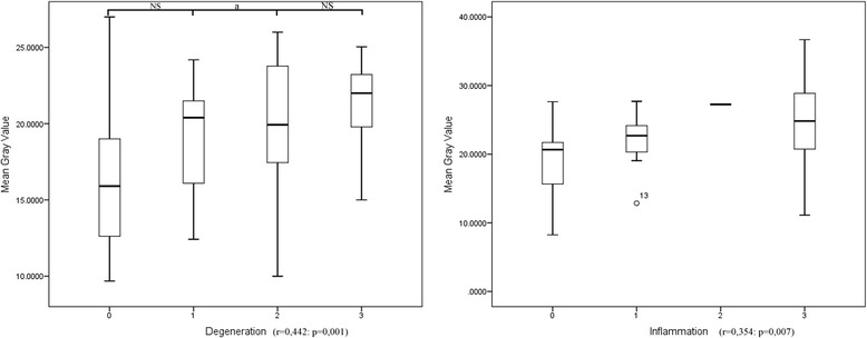
Results: Kidney samples were collected from 68 dog and 32 cat cadavers donated by the owners to the Veterinary Teaching Hospital of the University of Padua and standardized ultrasonographic images of each sample were collected. The echogenicity of the renal cortex was quantitatively assessed by means of mean gray value (MGV), and then histopathological analysis was performed. Statistical analysis to evaluate the influence of histological lesions on MGV was performed. The differentiation efficiency of MGV to detect pathological changes in the kidneys was calculated for dogs and cats. Statistical analysis revealed that only glomerulosclerosis was an independent determinant of echogenicity in dogs whereas interstitial nephritis, interstitial necrosis and fibrosis were independent determinants of echogenicity in cats. The global influence of histological lesions on renal echogenicity was higher in cats (23%) than in dogs (12%).
Conclusions: Different histopathological lesions influence the echogenicity of the kidneys in dogs and cats. Moreover, MGV is a poor test for distinguishing between normal and pathological kidneys in the dog with a sensitivity of 58.3% and specificity of 59.8%. Instead, it seems to perform globally better in the cat, resulting in a fair test, with a sensitivity of 80.6% and a specificity of 56%.
Figures


References
-
- d’Anjou MA. Kidneys and Ureters. In: Pennink D, d’Anjou MA, editors. Atlas of small animal ultrasonography. Ames, Iowa: Blackwell Publishing; 2008. pp. 339–64.
MeSH terms
LinkOut - more resources
Full Text Sources
Other Literature Sources
Medical
Miscellaneous
